Overview of Chest MRI
Magnetic Resonance Imaging (MRI) of the chest is a non-invasive diagnostic tool that utilizes a powerful magnetic field, radio waves, and a computer to generate detailed images of the structures within the chest cavity. Unlike X-rays and CT scans, MRI does not employ ionizing radiation, making it a safe and preferred option for many patients.
The high-resolution images produced by MRI provide a comprehensive view of the chest's internal anatomy, including the mediastinum (the central compartment housing the heart, major blood vessels, trachea, and esophagus), the chest wall, pleura (membranes surrounding the lungs), heart, and blood vessels. MRI offers the advantage of capturing images from virtually any angle, providing a thorough assessment of these structures. Furthermore, MRI can produce dynamic, movie-like sequences of the cardiovascular system, enabling doctors to evaluate the health and function of the heart, valves, and major blood vessels. This detailed information is invaluable for diagnosing and monitoring a wide range of chest conditions, from cardiovascular diseases to tumors and infections.
What are some common uses of the procedure?
MRI of the chest serves as a valuable tool for characterizing abnormal masses that may be difficult to assess with other imaging techniques like CT scans. It provides detailed information on tumor size, extent, and the degree of spread to nearby structures, aiding in accurate diagnosis and treatment planning.
Additionally, MRI is instrumental in evaluating the anatomy and function of the heart and its components, such as valves and chambers. It can assess myocardial perfusion (blood flow to the heart muscle), detect myocardial infarctions (scarring caused by prior blockages), and determine blood flow dynamics within the heart and blood vessels.
MRI is also used to visualize lymph nodes and blood vessels in the chest, helping to identify vascular and lymphatic malformations. It's helpful in assessing disorders of the chest bones (vertebrae, ribs, and sternum) and the soft tissues of the chest wall (muscles and fat). Moreover, it can detect and characterize pericardial disease, which affects the thin sac surrounding the heart.
When other imaging modalities like chest X-rays or CT scans detect lesions in the mediastinum (the central chest cavity) or pleura (the membranes surrounding the lungs), MRI can provide further clarification and detailed characterization. A specialized form of MRI, known as magnetic resonance angiography (MRA), is particularly useful for assessing the arteries and veins in the chest cavity. MRA can reveal aneurysms (abnormal bulges in blood vessels), thrombosis (blood clots), and dissections (tears in the inner lining of arteries).
How should I prepare for the Chest MRI?
Preparing for a chest MRI involves several steps to ensure a safe and successful procedure. You'll be asked to change into a hospital gown to avoid any artifacts in the images and comply with safety protocols related to the strong magnetic field. While typically you can eat and take medications as usual, specific instructions regarding food and drink might vary, so it's best to consult your doctor or the imaging center beforehand.
Some MRI exams utilize a contrast material called gadolinium to enhance image clarity. If your exam requires contrast, you'll be asked about any allergies or conditions like asthma, as well as allergies to medications, food, or environmental factors. Gadolinium is generally safe, with a lower risk of allergic reactions compared to iodine-based contrast. However, if you have a known allergy to gadolinium, pre-medication might be an option. Consult the ACR Manual on Contrast Media for more information.
It's crucial to inform the technologist or radiologist about any serious health conditions or recent surgeries. Certain conditions, like severe kidney disease, might require precautions when using gadolinium. A blood test may be necessary to assess kidney function.
Pregnant women should always inform their doctor and the technologist, as MRI is generally safe but requires special considerations during the first trimester. Gadolinium contrast is typically avoided unless absolutely necessary. For more information, refer to the MRI Safety During Pregnancy page.
If you experience claustrophobia or anxiety, discuss the possibility of a mild sedative with your doctor before the exam. Infants and young children often need sedation or anesthesia to remain still during the scan. The necessity depends on age, development, and the type of exam. Pediatric sedation specialists are available at many facilities to ensure their safety.
To create a comfortable experience for children, some facilities have staff trained to work with them, utilizing model scanners and explaining the procedure to alleviate anxiety. Goggles or headsets might be provided to watch movies during the scan, helping children stay still for optimal image quality.
Before entering the MRI room, remove all jewelry, accessories, and any metal or electronic items. These can interfere with the magnetic field, potentially causing damage or becoming projectiles. This includes items like jewelry, watches, credit cards, hearing aids, hairpins, metal zippers, removable dental work, pens, pocketknives, eyeglasses, body piercings, and electronic devices like mobile phones and smartwatches.
Most metal implants are safe in MRI, but exceptions include certain cochlear implants, aneurysm clips, metal coils in blood vessels, older cardiac devices, and vagal nerve stimulators. Inform the technologist of any implanted devices and provide any relevant documentation. If needed, an X-ray can be used to identify metal objects.
If you have shrapnel, bullets, or other metal fragments in your body, especially near the eyes, inform the medical staff. These can be hazardous due to potential movement or heating during the scan. While tattoo dyes containing iron might rarely cause issues, most dental fillings, braces, and cosmetics are generally safe, though they may affect image quality in the facial or brain areas. Finally, anyone accompanying you into the MRI room will also be screened for metal and electronic devices to ensure a safe environment.
What does the equipment look like?
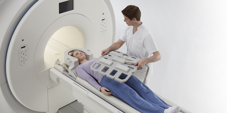
The typical MRI machine used for chest imaging is a large, cylindrical tube surrounded by a circular magnet. You'll lie on a movable table that slides into the center of this tube, placing you within the magnetic field for optimal imaging. However, alternative designs exist to accommodate varying patient needs and comfort levels.
Short-bore systems offer a less enclosed experience, with the magnet not fully surrounding you. Newer MRI machines often have wider bores, providing more space and comfort for larger individuals or those who experience claustrophobia. Additionally, "open" MRI units, with their open sides, offer a significantly less confined environment, making them particularly suitable for patients with anxiety related to enclosed spaces or those with larger body sizes.
While open MRI machines can produce high-quality images for many types of exams, there might be limitations for specific procedures. To determine the most suitable MRI unit for your specific needs, it's always best to consult with your radiologist, who can offer personalized guidance and address any concerns you may have.
How does the procedure work?
Unlike X-ray and computed tomography (CT) scans, which rely on ionizing radiation, MRI employs a powerful magnetic field and radio waves to create detailed images of the body's internal structures. This non-invasive technique works by temporarily realigning the hydrogen atoms naturally present in the body's tissues. As these atoms return to their normal alignment, they emit varying amounts of energy, depending on the type of tissue they are in.
The MRI scanner, equipped with coils that transmit and receive radio waves, captures these energy emissions and translates them into detailed visual representations. The magnetic field is generated by passing an electric current through wire coils within the machine, and additional coils may be placed around the specific body part being imaged. Importantly, the electric current itself does not come into contact with the patient.
A sophisticated computer then processes the signals emitted by the hydrogen atoms, creating a series of cross-sectional images, each depicting a thin slice of the body. These images can be viewed from different angles, allowing the radiologist to examine the internal structures in detail. MRI's exceptional ability to differentiate between healthy and diseased tissue makes it a valuable diagnostic tool, often surpassing the capabilities of X-ray, CT, and ultrasound in certain situations.
How is the procedure performed?
MRI exams of the chest are typically conducted on an outpatient basis, requiring no hospital stay. During the procedure, you will be comfortably positioned on a moveable table, and the technologist may use straps and bolsters to help you maintain a still position throughout the scan. Specialized devices containing coils that send and receive radio waves may be placed around or near your chest to capture detailed images.
The exam itself consists of multiple runs, or sequences, each lasting several minutes and producing distinct sounds. If your exam requires contrast material, a doctor, nurse, or technologist will insert an intravenous catheter (IV) into a vein in your hand or arm. This IV is used to administer the contrast material after an initial set of scans is completed.
Once you are positioned within the MRI machine, the technologist will operate the scanner from a nearby computer, monitoring the images as they are generated. You'll be able to communicate with the technologist through an intercom throughout the exam. If contrast material is used, additional images will be taken after the injection.
Upon completion of the scan, you might be asked to wait briefly while the radiologist reviews the images to ensure they are of sufficient quality and detail. The technologist will then remove the IV line, if applicable, and apply a small dressing to the insertion site.
The entire MRI exam usually takes about an hour, but the duration can vary depending on the complexity of the study and whether contrast material is used. If contrast is administered, the total scan time may be extended by 15 to 20 minutes.
What will I experience during and after the procedure?
While chest MRI exams are generally painless, some individuals may find it uncomfortable to remain still for extended periods. Additionally, the enclosed space within the MRI machine can trigger feelings of claustrophobia in certain patients. The scanner itself produces noise, but earplugs or headphones are provided to minimize discomfort.
It's normal for the imaged area to feel slightly warm due to the radiofrequency pulses, and if this becomes bothersome, informing the technologist is recommended. Maintaining stillness is crucial for obtaining clear images, and you'll know when images are being recorded due to the loud tapping or thumping sounds from the activated coils. Although you may be able to relax between sequences, maintaining your position is essential.
Despite being alone in the exam room, you'll be under constant observation and in communication with the technologist through a two-way intercom. A "squeeze-ball" is provided for immediate assistance if needed. Some facilities allow a screened companion to stay with you during the scan for added comfort.
For children undergoing the exam, appropriately sized earplugs or headphones are provided, and music might be played to help them relax and pass the time. MRI scanners are well-lit and air-conditioned to ensure a comfortable environment.
In some cases, a contrast material might be injected intravenously (IV) before the scan to enhance image clarity. The IV needle may cause slight discomfort or bruising, with a minimal risk of skin irritation at the insertion site. Some patients experience a temporary metallic taste after the injection.
If you don't require sedation, you can typically resume your normal activities and diet immediately after the exam. Although rare, some individuals may experience side effects like nausea, headache, or pain at the injection site. Allergic reactions, such as hives or itchy eyes, are exceedingly uncommon, but if they occur, the technologist should be notified immediately, and medical assistance will be provided promptly.
Who interprets the results and how do I get them?
A radiologist, a physician specializing in the interpretation of medical images, will meticulously analyze the MRI scans of your chest. After their thorough evaluation, a detailed report will be sent to your primary care physician or the doctor who referred you for the MRI. Your doctor will then discuss the results with you, explaining any findings or abnormalities in detail and outlining any necessary next steps.
In some cases, a follow-up exam may be recommended to gain a more comprehensive understanding of a potential issue, monitor its progression over time, or assess the effectiveness of any ongoing treatment. These follow-up scans could involve additional views, specialized imaging techniques, or other diagnostic tests. They are often crucial for ensuring accurate diagnosis and effective management of any chest conditions that may be identified during the initial MRI.
Benefits
Magnetic Resonance Imaging (MRI) of the chest is a non-invasive diagnostic technique that provides a wealth of information without exposing patients to radiation. Its ability to generate exceptionally clear and detailed images of the heart and vascular structures surpasses many other imaging modalities, making it invaluable for early detection and evaluation of cardiovascular conditions.
MRI's versatility extends beyond the heart and blood vessels, proving valuable in diagnosing a wide array of chest-related conditions, including bone and soft tissue abnormalities, as well as tumors. It allows physicians to assess not only the structure of organs but also their function, providing a comprehensive understanding of the chest's overall health.
MRI's ability to penetrate bone and visualize structures that might be hidden by bone in other imaging techniques makes it a powerful tool for identifying subtle abnormalities. Additionally, the gadolinium-based contrast material used in some MRI scans carries a lower risk of allergic reactions compared to iodine-based contrasts used in X-rays and CT scans.
While chest CT scans remain the preferred method for evaluating lung abnormalities, MRI often outperforms other imaging procedures in differentiating and characterizing soft tissues within the chest. Moreover, MRI can assess blood flow without the risks associated with traditional catheter angiography, making it a safer and less invasive alternative.
Risks
Chest MRI exams are generally considered safe for most individuals when appropriate safety guidelines are followed. However, certain risks should be acknowledged. While the use of sedation carries a small risk of over-sedation, vigilant monitoring of vital signs helps minimize this concern.
The strong magnetic field used in MRI poses no direct harm, but it can potentially interfere with the function of implanted medical devices or distort the resulting images. A rare complication associated with gadolinium-based contrast agents is nephrogenic systemic fibrosis, primarily observed in patients with pre-existing kidney issues. To mitigate this risk, doctors carefully assess kidney function before considering a contrast injection.
Allergic reactions to the contrast material are also possible, although they are typically mild and can be managed with medication. If you experience an allergic reaction, medical professionals will be readily available to provide immediate assistance. Recent research suggests that trace amounts of gadolinium might remain in the body, particularly in the brain, after multiple MRI scans. This is more likely to happen in patients who undergo frequent MRI exams over their lifetime for monitoring chronic or high-risk health conditions. Gadolinium is mainly excreted through the kidneys, but if you fall into this category, discuss the possibility of gadolinium retention with your doctor, as the effects can vary from person to person.
For breastfeeding mothers, manufacturers of IV contrast advise a 24-48 hour pause in breastfeeding after contrast administration. However, the latest guidance from the American College of Radiology indicates that the amount of contrast absorbed by infants through breast milk is minimal. For detailed information and guidance, consult the ACR Manual on Contrast Media and its references.
What are the limitations of a Chest MRI?
While MRI of the chest offers valuable diagnostic insights, it's important to be aware of its limitations. The quality of the images obtained relies heavily on your ability to remain perfectly still and follow breath-holding instructions during the scan. If you experience anxiety, confusion, or severe pain, maintaining stillness can be challenging, potentially compromising image clarity.
Body size can also be a constraint, as some MRI machines have weight limitations. Metal implants or other metallic objects in your body, as well as any movement during the procedure, can interfere with obtaining clear images. An irregular heartbeat might also affect image quality, particularly in techniques that rely on the heart's electrical activity for timing.
MRI is generally not recommended for critically injured patients due to potential interference from traction devices and life support equipment. However, medical professionals may still opt for MRI in certain trauma cases based on individual circumstances.
While non-contrast MRI is considered safe for pregnant women, doctors might postpone the scan until after delivery unless it's urgent. Gadolinium contrast agents are typically avoided during pregnancy except in specific circumstances. Your doctor will discuss the benefits and risks of any MRI procedure with you, and MRI might be performed after the first trimester to assess fetal development when ultrasound findings are inconclusive.
Compared to other imaging exams like X-rays or CT scans, MRI of the chest tends to be more time-consuming and expensive. If cost is a concern, it's advisable to consult with your insurance provider. Additionally, the longer duration of chest MRI exams often necessitates sedation for young children and infants to ensure they remain still throughout the procedure.