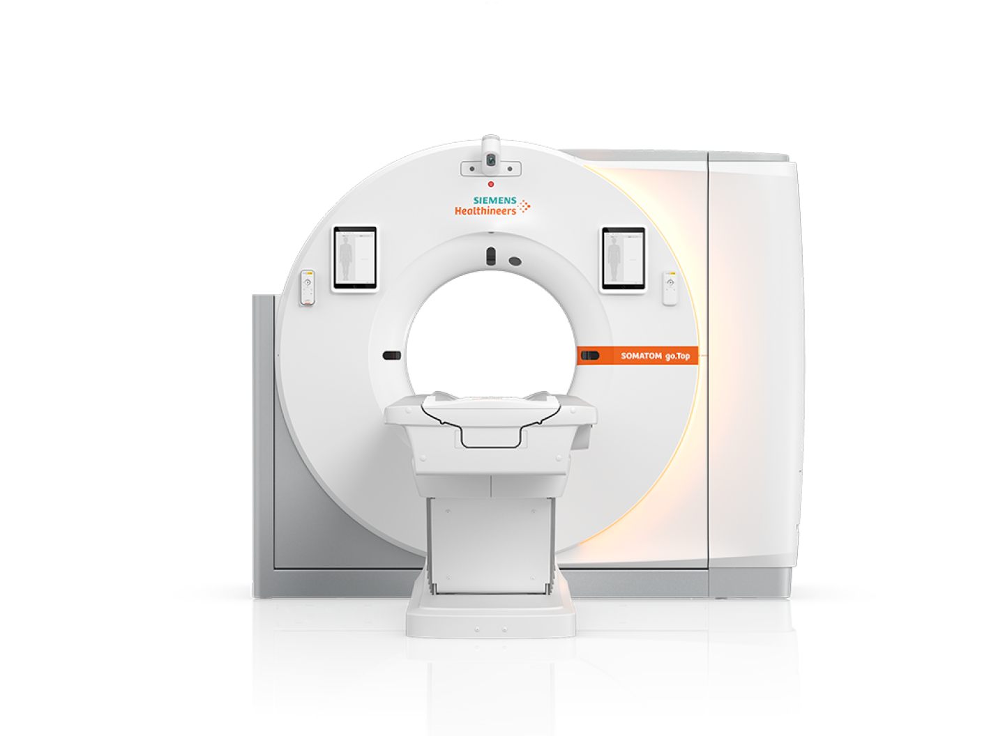
Overview of CT Scan
Computed tomography (CT) scanning is a powerful imaging technique that uses X-rays to create detailed, cross-sectional images of the body. Imagine slicing a loaf of bread—CT scans produce similar "slices" of your internal structures, allowing doctors to see bone, blood vessels, and soft tissues in incredible detail. This level of clarity surpasses that of traditional X-rays, providing a more comprehensive view of your anatomy.
During a CT scan, a special X-ray tube rotates around you while a table moves you through the scanner. A computer combines these X-ray images to create detailed 3D images of the area being examined. This allows doctors to see problems that might be missed with a standard X-ray, making CT scans invaluable for diagnosing a wide range of conditions, including injuries, tumors, infections, and blood vessel abnormalities.
Why it's done
Your healthcare professional might recommend a CT scan for a variety of reasons, providing valuable insights into your health. CT scans can help diagnose muscle and bone conditions, such as bone tumors and fractures. They can pinpoint the location of tumors, infections, or blood clots, and serve as a vital guide during procedures like surgery, biopsies, and radiation therapy.
CT scans are also instrumental in finding and monitoring the progress of various diseases and conditions, including cancer, heart disease, lung nodules, and liver masses. They can track the effectiveness of treatments, such as cancer therapies, and help identify internal injuries or bleeding that may occur after trauma.
The detailed images provided by CT scans allow doctors to make more informed diagnoses and treatment plans, leading to better outcomes for patients.
Risks
Radiation Exposure
While CT scans provide incredibly detailed images of your body, they do involve exposure to a type of energy called ionizing radiation. The amount of radiation used in a CT scan is greater than that of a standard X-ray because the scan gathers much more detailed information.
While the low doses of radiation used in CT scans haven't been shown to cause long-term harm, there might be a slight increase in the lifetime risk of cancer with repeated scans. This risk is generally considered small, especially in adults. However, children are more susceptible to the effects of radiation, so healthcare professionals carefully weigh the potential benefits against the risks for younger patients.
Despite this small risk, the numerous benefits of CT scans often outweigh them. Healthcare professionals are committed to using the lowest possible dose of radiation to obtain the necessary medical information. Moreover, newer, faster CT machines and techniques have significantly reduced radiation exposure compared to older models.
It's essential to discuss your concerns about radiation exposure with your healthcare provider. They can explain the benefits and risks of a CT scan in detail, helping you make an informed decision about your care.
Harm to Unborn Babies
If you are pregnant, it's crucial to inform your healthcare professional before undergoing a CT scan. While the radiation from a CT scan is unlikely to harm your baby unless the scan is specifically of your belly or pelvis, your doctor may suggest an alternative imaging method to avoid any potential exposure. Ultrasound and MRI are excellent alternatives that don't involve radiation and provide valuable insights into your health. Always prioritize open communication with your doctor about your pregnancy and any concerns you have regarding medical procedures. They will work with you to ensure the safest and most effective care for both you and your baby.
Contrast Material
For some CT scans, a special dye called contrast material is used to enhance the images. This dye appears bright on the scans, making it easier to see certain areas of the body, such as blood vessels, intestines, or other structures.
The contrast material can be administered in a few different ways:
By Mouth: If your esophagus or stomach is being scanned, you might be asked to drink a liquid containing contrast material. This liquid might not taste pleasant, but it helps the organs show up clearly on the CT scan.
By Injection: Contrast agents can be injected into an artery or vein in your arm. You might feel a brief sensation of warmth or a metallic taste in your mouth as the dye enters your system.
By Enema: In some cases, contrast material might be inserted into your rectum to help visualize your intestines. This procedure can cause a temporary feeling of bloating.
While contrast material is generally safe, it's important to be aware of potential complications, although they are rare. Most reactions to contrast material are mild, causing a rash or itchiness. However, in some cases, an allergic reaction can occur, and while uncommon, these can be serious, even life-threatening. If you've ever had a reaction to contrast material in the past, it's essential to inform your healthcare professional. Open communication with your doctor is vital to ensure a safe and comfortable CT scan experience.
How you Prepare
Before your CT scan, your healthcare provider might give you specific instructions depending on the area being examined. You might be asked to remove some or all of your clothing and wear a hospital gown. It's also important to remove any metal objects that could interfere with the scan, such as belts, jewelry, dentures, and eyeglasses. In some cases, you might need to fast for a few hours before the procedure to ensure an accurate scan. Your doctor will explain the necessary preparations in detail, ensuring a smooth and comfortable experience.
Preparing your Child for a Scan
If your infant or toddler is scheduled for a CT scan, your healthcare provider might recommend a sedative to help keep them calm and still during the procedure. Movement can blur the images, potentially affecting the results. It's essential for your child to remain as still as possible for an accurate scan. Your doctor can provide guidance on preparing your child for the CT scan. They may suggest ways to make the experience less stressful for your little one, such as bringing a favorite toy or blanket. Open communication with your healthcare provider is key to ensuring a positive and safe experience for your child.
What you can Expect
During the Test
During the CT scan procedure, you'll lie on a narrow table that slides into the center of a large, doughnut-shaped scanner. The table has a motor that moves you smoothly through the tunnel-like opening. You might be secured with straps and pillows to help you stay comfortable and still during the scan. For head scans, a special cradle may be used to hold your head steady.
As the table moves you through the scanner, the X-ray tube rotates around you, capturing images of thin slices of your body with each rotation. You may hear buzzing and whirring noises coming from the machine.
A CT technologist, a healthcare professional trained in operating the scanner, will monitor you from a separate room. You'll be able to communicate with them through an intercom if needed. To ensure clear images, the technologist might ask you to hold your breath at certain points during the scan. Movement can blur the images, making it difficult for the doctor to interpret the results.
Remember, the entire process is usually quick and painless. By following the technologist's instructions and remaining as still as possible, you'll help ensure a successful and accurate CT scan.
After the Test
After your CT scan, you can usually resume your regular activities. If you received contrast dye, you might be asked to wait for a short time before leaving to ensure you're feeling well. Your doctor might also recommend drinking plenty of fluids to help your kidneys flush the contrast material from your body.
The CT technologist will let you know when it's safe to leave and will provide any post-scan instructions. If you experience any unusual symptoms or have any concerns, be sure to contact your doctor.
Results
Your CT scan images are stored electronically and typically reviewed on a computer screen by a radiologist, a doctor who specializes in interpreting medical images. The radiologist will create a detailed report, which will be added to your medical records. Your healthcare provider will then review the report and discuss the findings with you, explaining what the images reveal and outlining any necessary next steps.