Overview of Shoulder MRI
Magnetic Resonance Imaging (MRI) of the shoulder is a non-invasive diagnostic technique that harnesses a powerful magnetic field and radio waves to create detailed, multi-dimensional images of the intricate structures within the shoulder joint. Unlike traditional X-rays, which primarily focus on bones, MRI offers a comprehensive view of the soft tissues, including tendons, muscles, ligaments, cartilage, and blood vessels. This comprehensive visualization enables healthcare professionals to accurately diagnose a wide range of shoulder conditions, from rotator cuff tears and labral injuries to arthritis and impingement syndromes. MRI's ability to detect subtle abnormalities and provide precise anatomical information makes it an invaluable tool in guiding treatment decisions and ensuring optimal outcomes for patients with shoulder pain or dysfunction.
What are some common uses of the procedure?
MRI of the shoulder excels in examining the intricate structures within the joint. It provides clear visualization of rotator cuff tears, injuries to the biceps tendon, and damage to the glenoid labrum, a crucial stabilizing component of the shoulder.
Common applications of shoulder MRI include diagnosing and evaluating degenerative joint disorders like arthritis and labral tears, as well as fractures in specific cases. It's particularly valuable for assessing rotator cuff disorders, including tears and impingement syndrome, which are major contributors to shoulder pain in individuals over 40.
MRI can also identify joint abnormalities caused by trauma, such as ligament and tendon tears. It's helpful in diagnosing sports-related injuries and work-related disorders stemming from repetitive strain, forceful impact, or vibration from hand-held tools. Additionally, it can detect infections like osteomyelitis and tumors affecting the bones, joints, or surrounding soft tissues.
Shoulder MRI is often used to investigate unexplained shoulder pain that doesn't improve with other treatments, decreased joint motion, and to monitor progress after shoulder surgery. In some cases, a specialized technique called magnetic resonance arthrography (MRA) may be employed, involving the injection of contrast material into the joint for enhanced visualization of specific structures.
How should I prepare for the Shoulder MRI?
Preparing for a shoulder MRI involves several key steps to ensure a safe and successful procedure. You will be asked to change into a hospital gown to eliminate any potential artifacts in the images and adhere to safety protocols due to the strong magnetic field. Guidelines regarding eating and drinking before the exam can vary, so it's important to consult your doctor or the imaging facility for specific instructions. Typically, you can continue your usual medication and diet unless advised otherwise.
Some shoulder MRI exams require the injection of a contrast material called gadolinium to enhance image clarity. Your doctor may inquire about any history of asthma or allergies to contrast materials, medications, food, or environmental factors. Gadolinium is generally safe, with a lower risk of allergic reactions compared to iodine-based contrast used in other imaging modalities. However, even with a known gadolinium allergy, it might be possible to use it with appropriate pre-medication. For detailed information on allergic reactions to gadolinium contrast, refer to the ACR Manual on Contrast Media.
It's crucial to inform the technologist or radiologist about any serious health conditions or recent surgeries, as these factors might influence the use of contrast or the overall safety of the procedure. Pregnant women should always inform their doctor and the technologist, as MRI is generally safe but requires special considerations during the first trimester. Gadolinium contrast is typically avoided unless absolutely necessary. For more information, refer to the MRI Safety During Pregnancy page.
If you experience claustrophobia or anxiety, discuss the possibility of a mild sedative with your doctor before the exam to alleviate any discomfort. Infants and young children often need sedation or anesthesia to remain still during the scan. The necessity depends on age, development, and the type of exam. Pediatric sedation specialists are available at many facilities to ensure their safety.
To create a comfortable experience for children, some facilities have staff trained to work with them, utilizing model scanners and explaining the procedure to alleviate anxiety. Goggles or headsets might be provided to watch movies during the scan, helping children stay still for optimal image quality.
Before entering the MRI room, remove all jewelry, accessories, and any metal or electronic items. These can interfere with the magnetic field, potentially causing damage to the equipment or becoming dangerous projectiles. This includes items like jewelry, watches, credit cards, hearing aids, hairpins, metal zippers, removable dental work, pens, pocketknives, eyeglasses, body piercings, and electronic devices like mobile phones and smartwatches.
Most metal implants are safe in MRI, but exceptions include certain cochlear implants, aneurysm clips, metal coils in blood vessels, older cardiac devices, and vagal nerve stimulators. Inform the technologist of any implanted devices and provide any relevant documentation to ensure safety during the scan. If needed, an X-ray can be used to identify metal objects.
If you have any shrapnel, bullets, or other metal fragments in your body, especially near the eyes, inform the medical staff. These can be hazardous due to potential movement or heating during the scan. While tattoo dyes containing iron might rarely cause issues, most dental fillings, braces, and cosmetics are generally safe, although they may affect image quality in the facial or brain areas. Finally, anyone accompanying you into the MRI room will also be screened for metal and electronic devices to ensure a safe environment.
What does the equipment look like?
The typical cardiac MRI machine is a large, cylindrical tube enclosed by a circular magnet, similar to other MRI units. During the scan, you'll lie on a movable table that slides into the center of this tunnel-like structure.
However, alternative designs exist to accommodate patient comfort and specific needs. Short-bore systems offer a more open configuration where the magnet doesn't completely surround you, providing a less confined experience. Additionally, newer MRI machines often feature larger diameter bores, allowing for more space and comfort, particularly for larger patients or those with claustrophobia.
"Open" MRI units represent a further departure from the traditional design, with open sides that create a significantly less claustrophobic environment. These units are particularly beneficial for individuals who experience anxiety in enclosed spaces or those with larger body sizes. While open MRI machines can produce high-quality images for many cardiac exams, their suitability for specific procedures may vary. It's advisable to consult with your radiologist to determine the most appropriate MRI unit for your individual needs.
How does the procedure work?
Unlike X-rays and computed tomography (CT) scans, which utilize ionizing radiation, MRI employs a powerful magnetic field and radio waves to create detailed images of the body's internal structures. This non-invasive technique works by temporarily realigning the hydrogen atoms naturally present within the body's tissues. As these atoms return to their normal alignment, they release varying amounts of energy, depending on the type of tissue they are in. This energy is detected by the scanner and used to construct a detailed image.
The MRI machine is equipped with coils that transmit and receive radio waves, which interact with the hydrogen atoms in the body. The magnetic field is generated by passing an electric current through wire coils within the machine, and additional coils may be placed around the specific body part being imaged. It's important to note that the electric current itself does not come into direct contact with the patient.
A sophisticated computer then processes the signals emitted by the hydrogen atoms, creating a series of cross-sectional images, each depicting a thin slice of the body. These images can be viewed from different angles, allowing the radiologist to examine the internal structures in detail. MRI's exceptional ability to differentiate between healthy and diseased tissue makes it a valuable diagnostic tool, often surpassing the capabilities of X-ray, CT, and ultrasound in certain situations.
How is the procedure performed?
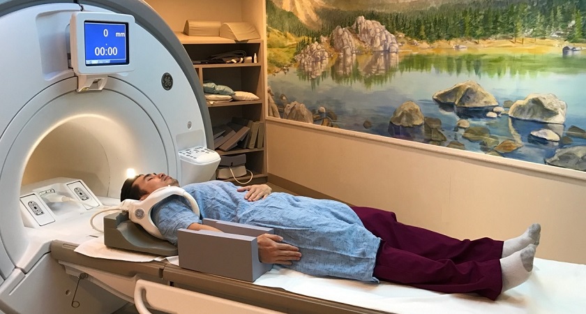
Shoulder MRI exams are usually outpatient procedures, meaning you can typically go home the same day. You'll be positioned comfortably on a movable table, with straps and bolsters potentially used to help you remain still during the scan. Small devices containing coils that send and receive radio waves may be placed around your shoulder to improve image quality.
If your exam involves contrast material, a doctor, nurse, or technologist will insert an intravenous catheter (IV) into a vein in your hand or arm for administration. After an initial series of scans, the contrast material will be injected, and additional images will be taken.
Once positioned within the MRI machine, the technologist will operate the scanner from a separate control room, but you'll be able to communicate with them through an intercom. After the scan, you might be asked to wait briefly while the radiologist reviews the images to ensure they are of sufficient quality. If an IV was used, it will be removed, and a small dressing will be applied.
The duration of the MRI exam can vary depending on the type of exam and the equipment used, but it typically takes between 15 and 45 minutes once imaging begins. If your child requires sedation, you may need to arrive earlier for a pre-sedation evaluation, and the procedure might take an additional 15 to 30 minutes. Your child may also need to be monitored afterward as the sedation wears off.
In some cases, a procedure called arthrography might be performed before the MRI. This involves injecting contrast material into the shoulder joint under image guidance to visualize the joint structures in greater detail.
What will I experience during and after the procedure?
While most MRI exams of the shoulder are painless, the need to remain still for extended periods can be uncomfortable for some individuals. Others may experience a sense of claustrophobia within the enclosed MRI scanner, and the machine itself can be noisy.
During the scan, it's normal to feel a slight warming sensation in the area being imaged due to the radiofrequency pulses. If this discomfort becomes bothersome, informing the technologist or radiologist is recommended. Maintaining stillness during image acquisition is crucial, as even slight movements can affect image quality. You'll be able to identify when images are being recorded by the loud tapping or thumping sounds produced by the activated coils. Although you may have opportunities to relax between sequences, maintaining your position is essential throughout the exam.
Despite being alone in the exam room, you will remain in constant contact with the technologist through a two-way intercom, allowing for communication throughout the procedure. A "squeeze-ball" will be provided, enabling you to alert the technologist immediately if you need assistance. Many facilities also permit a screened companion, such as a friend or family member, to stay with you during the scan for added comfort and support.
For children undergoing the exam, appropriately sized earplugs or headphones will be provided to minimize noise-related discomfort. Some facilities may even offer music through the headphones to help pass the time. MRI scanners are well-lit and air-conditioned to ensure a comfortable environment for all patients.
In some cases, an intravenous (IV) injection of contrast material may be administered before the scan to enhance image clarity. The IV needle can cause minor discomfort and bruising, and there's a slight risk of skin irritation at the insertion site. Some patients may experience a temporary metallic taste after the contrast injection.
If you haven't received sedation, there is no recovery period required after the MRI scan. You can immediately resume your usual activities, diet, and medications. While side effects from the contrast material are rare, some individuals may experience nausea, headache, or pain at the injection site. Allergic reactions, such as hives or itchy eyes, are extremely rare, but if they occur, inform the technologist so that immediate medical assistance can be provided.
Who interprets the results and how do I get them?
Following your shoulder MRI, a radiologist, a physician specializing in the interpretation of medical images, will carefully analyze the scans. They will then prepare a detailed report summarizing their findings and conclusions, which will be sent to the doctor who referred you for the MRI or your primary care physician. This doctor will discuss the results with you, explaining any identified abnormalities or issues in detail and outlining any necessary next steps, such as further testing or treatment options.
Benefits
MRI of the shoulder stands out as a non-invasive imaging technique, offering a radiation-free alternative for diagnosing a wide range of conditions. It's particularly valuable in identifying muscle and bone abnormalities within the shoulder joint. MRI can be instrumental in determining which patients with shoulder injuries require surgical intervention, providing crucial information for treatment decision-making. Additionally, it can help diagnose bone fractures when results from X-rays and other tests are inconclusive.
One of MRI's key advantages is its ability to detect abnormalities that might be obscured by bone in other imaging methods, offering a more comprehensive view of the shoulder's intricate structures. The gadolinium-based contrast material used in some MRI scans is less likely to cause allergic reactions than iodine-based contrasts used in X-rays and CT scans, making it a safer option for many patients.
Furthermore, MRI can serve as a non-invasive alternative to X-ray angiography and CT scans for diagnosing problems within the blood vessels of the shoulder. This eliminates the need for invasive procedures and reduces the associated risks, making it a preferred choice for many patients and healthcare providers.
Risks
Shoulder MRI scans are generally safe for most individuals when appropriate safety guidelines are followed. However, it's important to be aware of potential risks. While the use of sedation carries a small risk of over-sedation, vigilant monitoring of vital signs helps minimize this concern.
The strong magnetic field used in MRI poses no direct harm, but it can potentially interfere with the function of implanted medical devices or distort the resulting images. A rare complication associated with gadolinium-based contrast agents is nephrogenic systemic fibrosis, primarily observed in patients with pre-existing kidney issues. To mitigate this risk, doctors carefully assess kidney function before considering a contrast injection.
Allergic reactions to the contrast material are also possible, although they are typically mild and can be managed with medication. Medical assistance is readily available in case of an allergic reaction. Recent research suggests that trace amounts of gadolinium might remain in the body, particularly in the brain, after multiple MRI scans. This is more likely to happen in patients who undergo frequent MRI exams over their lifetime for monitoring chronic or high-risk health conditions. Gadolinium is mainly excreted through the kidneys, but if you fall into this category, discuss the possibility of gadolinium retention with your doctor, as the effects can vary from person to person.
For breastfeeding mothers, manufacturers of IV contrast advise a 24-48 hour pause in breastfeeding after contrast administration. However, the latest guidance from the American College of Radiology indicates that the amount of contrast absorbed by infants through breast milk is minimal. For detailed information and guidance, consult the ACR Manual on Contrast Media and its references.
What are the limitations of a Shoulder MRI?
While shoulder MRI offers exceptional diagnostic capabilities, it does have some limitations to consider. The quality of the images heavily relies on the patient's ability to remain perfectly still and follow breath-holding instructions during the scan. If you experience anxiety, confusion, or severe pain, maintaining stillness can be challenging, potentially affecting the clarity of the images.
Additionally, body size can be a limiting factor, as some MRI machines have weight restrictions, making it difficult for larger individuals to undergo the scan. The presence of metal implants or other metallic objects in the body can also interfere with image quality, as can patient movement during the procedure. In rare cases, a very irregular heartbeat might affect the quality of images due to the reliance of some MRI techniques on the heart's electrical activity for timing.
For pregnant women, non-contrast MRI is generally considered safe, with no conclusive evidence of harm to the fetus. However, doctors may opt to postpone the exam until after delivery unless it's deemed urgent. If an MRI is necessary during pregnancy, non-contrast scans are typically preferred after the first trimester to assess fetal development, while gadolinium contrast agents are generally avoided unless absolutely necessary. Your doctor will discuss the potential benefits and risks of any MRI procedure with you.
Finally, MRI exams tend to be more costly and time-consuming compared to other imaging techniques like X-rays or ultrasounds. If cost is a concern, it's advisable to discuss it with your insurance provider.