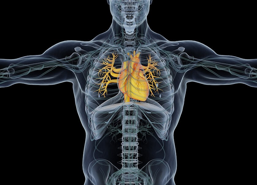
Overview of Cardiac MRI
Cardiac magnetic resonance imaging (MRI) is a non-invasive diagnostic technique that utilizes a powerful magnetic field, radio waves, and a computer to generate detailed images of the heart and its surrounding structures. This versatile tool is employed by doctors to detect, diagnose, and monitor a wide range of cardiac diseases, including both congenital heart conditions (present at birth) and those that develop later in life.
Unlike some other imaging methods, cardiac MRI does not involve exposure to ionizing radiation, making it a safe option for patients. It is particularly valuable for certain conditions where it can provide superior image quality compared to other modalities, offering detailed visualization of the heart's anatomy and function. This detailed information aids physicians in making accurate diagnoses, developing personalized treatment plans, and monitoring the progression of heart diseases over time.
What are some common uses of the procedure?
Cardiac MRI serves as a versatile tool for diagnosing and monitoring a wide range of heart conditions. It allows doctors to assess the anatomy and function of the heart chambers, valves, major blood vessels, and surrounding structures like the pericardium. This comprehensive evaluation aids in diagnosing various cardiovascular disorders, including tumors, infections, and inflammatory conditions.
MRI is particularly valuable in assessing the impact of coronary artery disease, revealing the extent of limited blood flow to the heart muscle and scarring caused by heart attacks. It also helps in planning and monitoring the progression of treatments for various cardiovascular disorders, especially in patients with congenital heart disease.
Furthermore, cardiac MRI is instrumental in evaluating the heart's anatomy and function in individuals with both congenital and acquired heart diseases. It aids in the pre-operative planning and post-operative monitoring of surgical interventions for congenital heart defects.
Beyond these applications, cardiac MRI is used to diagnose and monitor the progression or recovery from heart failure and arrhythmias, assess pericardial diseases, evaluate heart valve function and regurgitation, monitor the size of major blood vessels like the aorta, and evaluate masses in or around the heart. It is also essential in the diagnosis and management of hypertrophic cardiomyopathy, a condition characterized by the overgrowth of portions of the heart muscle.
How should I prepare for the Cardiac MRI?
Preparing for a cardiac MRI involves several important steps to ensure a safe and successful procedure. You'll be asked to change into a hospital gown to avoid any artifacts in the images and comply with safety protocols related to the strong magnetic field. Typically, you can continue your usual medication and diet unless your doctor advises otherwise, but guidelines regarding food and drink may vary depending on the specific exam and facility.
Some cardiac MRI exams involve the injection of a contrast material called gadolinium to enhance image clarity. Your doctor will inquire about any history of asthma or allergies to contrast materials, medications, food, or environmental factors. Gadolinium is generally safe, with a lower risk of allergic reactions compared to iodine-based contrast used in other imaging modalities. However, even with a known gadolinium allergy, it might be possible to use it with appropriate pre-medication. For detailed information on allergic reactions to gadolinium contrast, refer to the ACR Manual on Contrast Media.
It's crucial to inform the technologist or radiologist about any serious health conditions or recent surgeries, as these factors might influence the use of contrast or the overall safety of the procedure. Pregnant women should always inform their doctor and the technologist, as MRI is generally safe but requires special considerations during the first trimester. Gadolinium contrast is typically avoided unless absolutely necessary. For more information, refer to the MRI Safety During Pregnancy page.
If you experience claustrophobia or anxiety, discuss the possibility of a mild sedative with your doctor before the exam to alleviate any discomfort. Infants and young children often need sedation or anesthesia to remain still during the scan. The necessity depends on age, development, and the type of exam. Pediatric sedation specialists are available at many facilities to ensure their safety.
To create a comfortable experience for children, some facilities have staff trained to work with them, utilizing model scanners and explaining the procedure to alleviate anxiety. Goggles or headsets might be provided to watch movies during the scan, helping children stay still for optimal image quality.
Before entering the MRI room, remove all jewelry, accessories, and any metal or electronic items. These can interfere with the magnetic field, potentially causing damage to the equipment or becoming dangerous projectiles. This includes items like jewelry, watches, credit cards, hearing aids, hairpins, metal zippers, removable dental work, pens, pocketknives, eyeglasses, body piercings, and electronic devices like mobile phones and smartwatches.
Most metal implants are safe in MRI, but exceptions include certain cochlear implants, aneurysm clips, metal coils in blood vessels, older cardiac devices, and vagal nerve stimulators. Inform the technologist of any implanted devices and provide any relevant documentation to ensure safety during the scan. If needed, an X-ray can be used to identify metal objects.
If you have any shrapnel, bullets, or other metal fragments in your body, especially near the eyes, inform the medical staff. These can be hazardous due to potential movement or heating during the scan. While tattoo dyes containing iron might rarely cause issues, most dental fillings, braces, and cosmetics are generally safe, although they may affect image quality in the facial or brain areas. Finally, anyone accompanying you into the MRI room will also be screened for metal and electronic devices to ensure a safe environment.
What does the equipment look like?
The typical cardiac MRI machine is a large, cylindrical tube enclosed by a circular magnet, similar to other MRI units. During the scan, you'll lie on a movable table that slides into the center of this tunnel-like structure.
However, alternative designs exist to accommodate patient comfort and specific needs. Short-bore systems offer a more open configuration where the magnet doesn't completely surround you, providing a less confined experience. Additionally, newer MRI machines often feature larger diameter bores, allowing for more space and comfort, particularly for larger patients or those with claustrophobia.
"Open" MRI units represent a further departure from the traditional design, with open sides that create a significantly less claustrophobic environment. These units are particularly beneficial for individuals who experience anxiety in enclosed spaces or those with larger body sizes. While open MRI machines can produce high-quality images for many cardiac exams, their suitability for specific procedures may vary. It's advisable to consult with your radiologist to determine the most appropriate MRI unit for your individual needs.
How does the procedure work?
Unlike X-rays and computed tomography (CT) scans, which rely on ionizing radiation, MRI employs a powerful magnetic field and radio waves to create detailed images of the body's internal structures. This non-invasive technique works by temporarily realigning the hydrogen atoms naturally present within the body's tissues. As these atoms return to their normal alignment, they emit different amounts of energy depending on the type of tissue they reside in. The scanner detects and measures these energy variations, using this information to construct a detailed image of the heart and surrounding areas.
The MRI machine is equipped with coils that transmit and receive radio waves, which interact with the hydrogen atoms in the body. The magnetic field is generated by passing an electric current through wire coils within the machine, and additional coils may be placed around the chest area to enhance image acquisition. It's important to note that the electric current itself does not come into direct contact with the patient.
A sophisticated computer then processes the signals emitted by the hydrogen atoms, creating a series of cross-sectional images, each depicting a thin slice of the chest. These images can be viewed from various angles, providing a comprehensive and detailed view of the heart's structure and function. MRI's exceptional ability to differentiate between healthy and diseased tissue makes it a valuable diagnostic tool, often surpassing the capabilities of X-ray, CT, and ultrasound in assessing cardiac conditions.
How is the procedure performed?
Cardiac MRI exams are typically performed on an outpatient basis, and you will be positioned on a movable table by the technologist, who may use straps and bolsters to ensure you remain still during the scan. Specialized coils that send and receive radio waves will be placed around or near your chest to capture detailed images.
The exam involves multiple sequences, each lasting several minutes and producing distinct sounds. To synchronize image acquisition with your heartbeat, electrocardiogram (ECG) leads (sticky patches) will be placed on your chest, and a respiratory gating belt might be placed around your abdomen to monitor your breathing. A pulse monitor on your finger will also be used. You'll be given breathing instructions and may be asked to hold your breath briefly during certain sequences.
If your exam requires contrast material, an intravenous catheter (IV) will be inserted into a vein in your hand or arm for administration. After an initial series of scans, the technologist will inject the contrast material through the IV and take additional images during or following the injection.
Once inside the MRI machine, the technologist will operate the scanner from a separate room, but you'll be able to communicate with them through an intercom. After the scan, you might be asked to wait briefly while the radiologist reviews the images. The IV line will be removed, and a small dressing applied if necessary.
The entire exam, including preparation and imaging, usually takes 90 minutes or less. However, the duration can vary depending on the complexity of the study and the findings observed during the scan. If your child is undergoing the exam under sedation or anesthesia, the recovery period typically ranges from 30 minutes to two hours.
What will I experience during and after the procedure?
While cardiac MRI exams are generally painless, some individuals may find it challenging to remain still for extended periods. Additionally, the enclosed space within the MRI machine can trigger feelings of claustrophobia in certain patients. The scanner itself generates noise, but earplugs or headphones are provided to minimize discomfort.
During the exam, the technologist will closely monitor your heartbeat and guide you through brief breath-holding exercises to optimize image quality. It's normal for the imaged area to feel slightly warm due to the radiofrequency pulses, but if it becomes bothersome, informing the technologist is recommended. Maintaining stillness throughout the scan is crucial, particularly during the few seconds to minutes when images are being recorded. The loud tapping or thumping sounds you'll hear and feel are simply the coils generating the radio waves. While you may be able to relax between imaging sequences, maintaining your position as much as possible is important.
Throughout the exam, you won't be alone, as the technologist can see, hear, and communicate with you through a two-way intercom. You'll also be given a "squeeze-ball" to alert them if you need immediate attention. Many facilities allow a screened companion to stay with you during the scan, providing an extra layer of comfort.
If contrast material is used, you might experience a brief sensation of coolness and flushing upon injection. The IV needle can cause minor discomfort and bruising, and there's a slight chance of skin irritation at the insertion site. Some patients also report a temporary metallic taste after the injection.
For most patients, there's no recovery period needed after the exam, and you can resume your usual activities and diet. While rare, side effects from the contrast material, such as nausea, headache, or pain at the injection site, can occur. Allergic reactions like hives or itchy eyes are exceedingly uncommon, but if they happen, the technologist should be alerted immediately for prompt medical attention.
Who interprets the results and how do I get them?
A radiologist, a physician specializing in interpreting medical images, will meticulously analyze the images obtained during your cardiac MRI. They will then compile a detailed report outlining their findings and conclusions, which will be sent to the doctor who referred you for the MRI or your primary care physician. Your doctor will then discuss the results with you, explaining any identified abnormalities or issues in detail and outlining any necessary next steps.
In some cases, a follow-up exam might be recommended to gain a deeper understanding of a potential issue, monitor any changes over time, or assess the effectiveness of an ongoing treatment plan. These follow-up scans could involve additional views, specialized imaging techniques, or other diagnostic tests. They are often crucial for ensuring accurate diagnosis and effective management of any cardiac conditions that may be identified during the initial MRI.
Benefits
Cardiac MRI stands out as a non-invasive diagnostic tool that offers numerous advantages in the assessment of heart health. Unlike many other imaging modalities, it doesn't expose patients to harmful radiation. For specific conditions, MRI provides superior image quality compared to other methods, making it an invaluable tool for early detection and evaluation of various cardiac abnormalities, particularly those affecting the heart muscle.
MRI has proven to be highly effective in diagnosing a wide range of cardiac conditions, including congenital heart defects, valve dysfunction, tumors, and diseases related to coronary artery disease and cardiomyopathy. Furthermore, it can be utilized during certain interventional procedures, such as catheter-based ablation for irregular heart rhythms, significantly reducing procedure time and enhancing accuracy.
MRI's ability to penetrate bone and visualize structures that might be obscured in other imaging techniques makes it a powerful tool for identifying subtle abnormalities. Additionally, the gadolinium-based contrast material used in some MRI scans carries a lower risk of allergic reactions compared to iodine-based contrasts used in X-rays and CT scans.
A key advantage of cardiac MRI is its ability to provide a comprehensive evaluation of both the structure and function of the heart and major blood vessels without exposing patients to the radiation risks associated with other imaging procedures. This makes it a safer and preferred option for many individuals, particularly those requiring repeated imaging or those who are more sensitive to radiation exposure.
Risks
Cardiac MRI exams are generally considered safe for most individuals when appropriate safety guidelines are followed. However, it's important to be aware of potential risks. While the use of sedation carries a small risk of over-sedation, vigilant monitoring of vital signs helps minimize this concern.
The strong magnetic field used in MRI poses no direct harm but can potentially interfere with the function of implanted medical devices or distort the resulting images. In rare cases, nephrogenic systemic fibrosis, a complication linked to gadolinium-based contrast agents, can occur, primarily in patients with pre-existing kidney issues. To mitigate this risk, doctors carefully assess kidney function before considering a contrast injection.
Allergic reactions to the contrast material are also possible, though they are typically mild and can be managed with medication. Medical assistance is readily available in case of an allergic reaction. Recent research suggests that trace amounts of gadolinium might remain in the body, particularly in the brain, after multiple MRI scans. This is more likely to happen in patients who undergo frequent MRI exams over their lifetime for monitoring chronic or high-risk health conditions. Gadolinium is mainly excreted through the kidneys, but if you fall into this category, discuss the possibility of gadolinium retention with your doctor, as the effects can vary from person to person.
For breastfeeding mothers, manufacturers of IV contrast advise a 24-48 hour pause in breastfeeding after contrast administration. However, the latest guidance from the American College of Radiology indicates that the amount of contrast absorbed by infants through breast milk is minimal. For detailed information and guidance, consult the ACR Manual on Contrast Media and its references.
What are the limitations of a Cardiac MRI?
Cardiac MRI, while a valuable diagnostic tool, has certain limitations. The quality of the images hinges on your ability to remain still and follow breath-holding instructions during the scan. Anxiety, confusion, or severe pain can make this difficult, potentially impacting image clarity. Additionally, some MRI machines have weight limits, posing challenges for larger individuals.
The presence of implants or other metallic objects in the body can also interfere with obtaining clear images, as can patient movement. Irregular heartbeats, particularly atrial fibrillation, can further complicate image acquisition due to the unpredictable nature of cardiac motion and heart rate. However, various techniques can overcome these challenges, such as synchronizing imaging with ECG or breathing patterns, or performing repeated breath-holds.
MRI is generally not recommended for critically injured patients due to potential interference from medical equipment. However, doctors may still consider MRI in specific trauma cases based on clinical judgment.
While non-contrast MRI is considered safe for pregnant women, doctors might postpone the exam if not urgent, as the strong magnetic field's effects on the fetus are still being studied. Gadolinium contrast agents are generally avoided during pregnancy unless absolutely necessary.
A notable limitation of cardiac MRI is the difficulty in obtaining detailed images of the coronary arteries and their branches compared to other imaging methods like cardiac CT or invasive angiography.
Lastly, cardiac MRI exams tend to be more expensive and time-consuming than other imaging techniques. If cost is a concern, discussing it with your insurance provider is recommended.