Overview of CT 3D - Face Scan
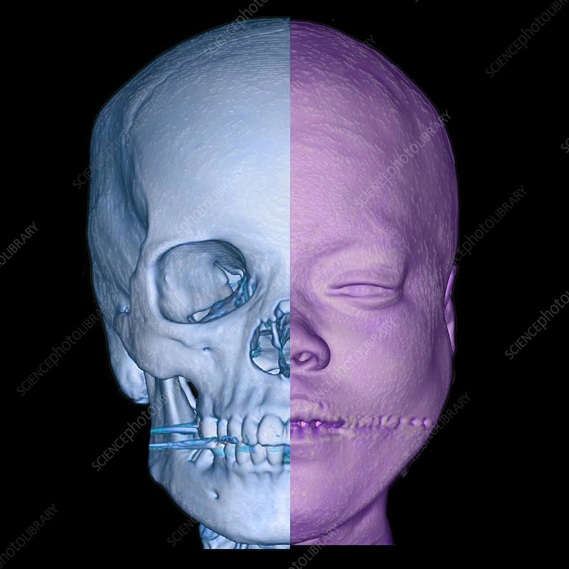
A 3D CT scan of the face provides detailed imaging to visualize the bony structures of the face in three dimensions. This advanced imaging technique uses computed tomography (CT) technology to create a comprehensive, three-dimensional representation of facial anatomy, including the skull, sinuses, nasal cavity, and facial bones.
The procedure involves taking multiple cross-sectional CT images of the face from various angles. These images are then reconstructed by specialized software to produce a detailed 3D model of the facial bones. This 3D model allows for an enhanced view of complex facial structures, which is particularly useful for assessing fractures, congenital anomalies, and other bony abnormalities.
3D CT face imaging is often employed in various clinical scenarios, such as planning reconstructive surgery or facial trauma repair. It provides surgeons with a precise and detailed map of the facial anatomy, facilitating accurate surgical planning and improved outcomes. It is also useful in evaluating and diagnosing conditions like facial deformities, sinus issues, and dental problems, and in forensic cases where detailed facial bone analysis is required.
The high level of detail provided by 3D CT face scans makes it a valuable tool in both diagnostic and preoperative settings, offering a clearer understanding of facial structures that traditional 2D imaging may not fully reveal.
What are some common uses of the procedure?
Doctors commonly recommend 3D CT face scans to assess and diagnose a variety of conditions related to the facial structure. This imaging technique is particularly effective in detecting facial fractures, including those of the orbital floor, which can occur due to trauma or injury. It provides a detailed view of the bony structures, making it easier to identify and evaluate fractures.
In addition to detecting fractures, 3D CT scans are used to identify and monitor tumors within the facial region. The detailed 3D reconstruction helps in distinguishing between benign and malignant growths and assessing their extent.
The scan is also instrumental in evaluating the extent of trauma to the facial bones and surrounding structures, which is crucial for planning appropriate treatment and surgical intervention. Furthermore, 3D CT face scans are useful in diagnosing sinusitis, particularly when it involves the bony structures of the nose and sinuses. This imaging modality provides a comprehensive view of the sinus cavities and their relationship with other facial structures, aiding in accurate diagnosis and treatment planning.
How should I prepare for a CT scan?
For a CT scan, wear comfortable, loose-fitting clothing. You may be required to change into a gown for the procedure. Metal objects, including jewelry, eyeglasses, dentures, and hairpins, can interfere with the CT images, so it is advisable to leave them at home or remove them before the exam. Some CT exams may also require the removal of hearing aids and removable dental work. Women should remove bras containing metal underwire, and you may need to remove any piercings if possible.
If your CT scan involves the use of contrast material, your doctor may instruct you to refrain from eating or drinking for a few hours before the exam. Inform your doctor about all medications you are taking and any allergies you have. If you have a known allergy to contrast material, your doctor may prescribe medications, usually a steroid, to reduce the risk of an allergic reaction. To avoid unnecessary delays, contact your doctor well in advance of the exam date.
Additionally, inform your doctor about any recent illnesses or other medical conditions, and whether you have a history of heart disease, asthma, diabetes, kidney disease, or thyroid problems, as these conditions may increase the risk of an adverse effect. The radiologist should also be informed if you have asthma, multiple myeloma, or any disorder of the heart, kidneys, or thyroid gland, or if you have diabetes, particularly if you are taking Metformin.
Women should always inform their physician and the CT technologist if there is any possibility that they may be pregnant. For more information, refer to the CT Safety During Pregnancy page.
What does the equipment look like?
The CT scanner is typically a large, donut-shaped machine with a short tunnel in the center. You will lie on a narrow table that slides in and out of this short tunnel. Surrounding you, the x-ray tube and electronic x-ray detectors are positioned opposite each other in a ring, called a gantry. The computer workstation that processes the imaging information is located in a separate control room. This is where the technologist operates the scanner and monitors your exam, maintaining direct visual contact with you. The technologist can hear and communicate with you using a speaker and microphone.
How does the procedure work?
A CT scan operates similarly to other x-ray exams, relying on the differential absorption of x-rays by various body parts. This difference allows doctors to distinguish between different body structures on an x-ray or CT image. During a conventional x-ray exam, a small amount of radiation is directed through the body part under examination, with a special electronic image recording plate capturing the resulting image. On an x-ray, bones appear white, soft tissues such as the heart or liver show up in shades of gray, and air appears black.
In contrast, CT scanning employs multiple x-ray beams and electronic x-ray detectors that rotate around the patient, measuring the radiation absorbed throughout the body. Occasionally, the exam table will move during the scan. A specialized computer program processes the large volume of data to create two-dimensional cross-sectional images of the body, which are then displayed on a computer monitor. CT imaging is often compared to slicing a loaf of bread into thin slices and then examining each slice; when the computer reassembles these image slices, it provides a highly detailed multidimensional view of the body's interior.
Most modern CT scanners can obtain multiple slices in a single rotation, known as multi-slice (multidetector) CT scanners, which capture thinner slices in less time, resulting in more detailed images. These advanced CT scanners can image large sections of the body in mere seconds, and even faster in small children. This speed benefits all patients, especially those who find it challenging to remain still, such as children, the elderly, and critically ill individuals.
For pediatric patients, radiologists adjust the CT scanner techniques to their size and the area of interest, thereby reducing the radiation dose. Some CT exams also use a contrast material to enhance the visibility of the body area under examination.
How is the procedure performed?
The technologist begins by positioning you on the CT exam table, typically lying flat on your back. Straps and pillows may be used to help you maintain the correct position and remain still during the exam. Many modern CT scanners are fast enough to scan children without the need for sedation; however, in special cases where children cannot hold still, sedation may be necessary. Motion during the scan can cause blurring of the images and degrade image quality, similar to how it affects photographs.
Depending on the type of exam, contrast material may be used. If required, the contrast material will be swallowed, injected through an intravenous line (IV), or, rarely, administered by enema. Once you are positioned, the table will move quickly through the scanner to determine the correct starting position for the scans. The table will then move slowly through the machine for the actual CT scan. Depending on the specific type of CT scan, the machine may make several passes.
The technologist may ask you to hold your breath during the scanning process. Any motion, including breathing and body movements, can lead to artifacts on the images, causing a loss of image quality that resembles the blurring seen in photographs of moving objects. When the exam is complete, the technologist will ask you to wait until they verify that the images are of high enough quality for accurate interpretation by the radiologist.
A CT scan of the usually takes about 10-45 minutes to complete.
What will I experience during and after the procedure?
CT exams are generally painless, fast, and easy, with multidetector CT reducing the amount of time you need to lie still. Although the scanning itself causes no pain, there may be some discomfort from having to remain still for several minutes. If you have difficulty staying still, are claustrophobic, or have chronic pain, you may find the CT exam stressful. The technologist or nurse, under the direction of a physician, may offer you medication to help you tolerate the procedure.
If the exam uses iodinated contrast material, your doctor will screen you for chronic or acute kidney disease. The contrast material may be administered intravenously, so you will feel a pin prick when the nurse inserts the needle into your vein. As the contrast is injected, you might feel warm or flushed, and you may experience a metallic taste in your mouth. These sensations are normal and will pass quickly. You might also feel a need to urinate, but this is only a side effect of the contrast injection and will subside rapidly.
Upon entering the CT scanner, you may see special light lines projected onto your body, which help ensure that you are in the correct position on the exam table. With modern CT scanners, you may hear slight buzzing, clicking, and whirring sounds as the internal parts of the CT scanner revolve around you during the imaging process.
During the CT scan, you will be alone in the exam room unless there are special circumstances, such as a parent wearing a lead shield staying in the room with their child. However, the technologist will always be able to see, hear, and speak with you through a built-in intercom system. For pediatric patients, a parent may be allowed in the room but may need to wear a lead apron to minimize radiation exposure.
After the CT exam, the technologist will remove your intravenous line and cover the tiny hole made by the needle with a small dressing. You can return to your normal activities immediately.
Who interprets the results and how do I get them?
A radiologist, a doctor specially trained to supervise and interpret radiology exams, will analyze the images from your CT scan. After analyzing the images, the radiologist will send an official report to the doctor who ordered the exam. Your doctor will then review the results with you and explain any findings.
In some cases, you may need a follow-up exam. If so, your doctor will explain the reason for this additional examination. A follow-up exam might be necessary to further evaluate a potential issue with more views or a special imaging technique, or to monitor any changes in a condition over time. Follow-up exams are often the best way to determine if treatment is effective or if a problem requires further attention.
Benefits
CT scanning offers numerous benefits, making it a valuable tool in medical imaging. It is a painless, noninvasive, and accurate procedure. One major advantage of CT scanning is its ability to image bone, soft tissue, and blood vessels simultaneously. Unlike conventional x-rays, CT scans provide highly detailed images of various types of tissue, including the lungs, bones, and blood vessels. The process is fast and simple, which is particularly beneficial in emergency cases where internal injuries and bleeding need to be identified quickly to help save lives.
CT scanning is also a cost-effective imaging tool for a wide range of clinical problems. It is less sensitive to patient movement compared to MRI, and unlike MRI, having an implanted medical device does not prevent you from undergoing a CT scan. Additionally, a diagnosis determined by CT scanning can eliminate the need for exploratory surgery and surgical biopsy.
No radiation remains in a patient's body after a CT exam, and the x-rays used for CT scanning should have no immediate side effects. These benefits highlight why CT scanning is a preferred choice in many medical scenarios.
Risks
There is always a slight risk of cancer associated with excessive exposure to radiation from CT scans. However, the benefit of obtaining an accurate diagnosis generally outweighs this risk. The radiation dose for CT procedures can vary; detailed information about radiation dose is available on the Radiation Dose page.
Women should inform their doctor and the CT technologist if there is any possibility of pregnancy, as CT scanning is generally not recommended for pregnant women unless absolutely necessary due to potential risks to the developing baby. The risk is minimal for head CT scans, but caution is advised. For more information on pregnancy and x-rays, refer to the Radiation Safety page.
If IV contrast material is used during the scan, manufacturers recommend that breastfeeding mothers avoid nursing for 24-48 hours afterward. However, the American College of Radiology (ACR) Manual on Contrast Media reports that the amount of contrast absorbed by an infant during breastfeeding is extremely low. For further details, consult the ACR Manual on Contrast Media and its references.
The risk of a serious allergic reaction to iodine-based contrast materials is extremely rare, and radiology departments are well-prepared to handle such reactions.
Children are more sensitive to radiation, so CT scans should be performed only when essential for diagnosis. They should not undergo repeated CT exams unless necessary, and CT scans in children should be conducted using low-dose techniques to minimize radiation exposure.
What are the limitations of 3D - Face CT?
Despite its advanced imaging capabilities, the 3D CT face scan has several limitations. One major limitation is its inability to capture detailed information about soft tissues, such as muscles and nerves, which are not well-visualized compared to bony structures. Consequently, it may not provide a complete assessment of soft tissue injuries or pathologies.
Another limitation is related to radiation exposure. While modern CT scanners are designed to minimize radiation doses, 3D CT scans still involve exposure to X-rays, which may be a concern for patients requiring multiple scans or those who are particularly sensitive to radiation.
The effectiveness of the scan can also be impacted by motion artifacts. If a patient is unable to remain still during the scan, the resulting images may be blurred or distorted, which can affect the accuracy of the diagnosis.
Finally, 3D CT face scans can be less effective for evaluating certain conditions, such as small or subtle bony abnormalities, compared to other imaging modalities like MRI or specialized X-ray techniques. In some cases, additional imaging or diagnostic methods may be necessary to obtain a comprehensive assessment of complex facial issues.