Overview of Breast MRI
Breast MRI, or magnetic resonance imaging of the breast, is a non-invasive diagnostic tool that utilizes a powerful magnetic field, radio waves, and a computer to generate highly detailed images of the breast's internal structures. While primarily used as a supplementary tool to mammography or ultrasound, breast MRI plays a crucial role in several scenarios. It can be used to screen women at high risk for breast cancer, assess the extent of cancer after a diagnosis, or further evaluate abnormalities detected on mammograms. One of its significant advantages is the absence of ionizing radiation, making it a safer option for repeated imaging. Additionally, breast MRI is considered the gold standard for determining whether silicone breast implants have ruptured.
Furthermore, breast MRI offers unique insights into various breast conditions that might not be readily apparent through other imaging modalities like mammography or ultrasound. This makes it a valuable tool in the comprehensive assessment of breast health, providing critical information to guide diagnosis and treatment decisions.
What are some common uses of the procedure?
Breast MRI, while not a replacement for mammography or ultrasound, is a valuable supplementary tool with several important applications. It can be used for screening women with a high risk of breast cancer, often due to a strong family history, providing an additional layer of detection alongside mammograms.
After a breast cancer diagnosis, MRI helps determine the extent of the disease, including tumor size, involvement of underlying muscle, the presence of additional tumors in the same or opposite breast, and any enlarged lymph nodes in the armpit, indicating potential spread.
When abnormalities detected on mammograms are difficult to assess with further mammography or ultrasound, MRI can offer a definitive evaluation, determining whether a biopsy is necessary or if the abnormality is benign.
For women who have undergone lumpectomies, MRI can distinguish between normal scar tissue and recurrent cancer, which can appear similar on mammograms and ultrasounds.
In cases where chemotherapy is administered before surgery (neoadjuvant chemotherapy), MRI monitors treatment progress and reassesses the remaining tumor size before surgical intervention.
Lastly, MRI is the most reliable method for detecting ruptures in silicone breast implants, ensuring the safety and integrity of these devices.
How should I prepare for the Breast MRI?
Preparing for a breast MRI involves several key steps to ensure a smooth and safe experience. You will be asked to change into a hospital gown to eliminate any potential artifacts in the images and adhere to safety protocols due to the strong magnetic field. Guidelines regarding eating and drinking before the exam can vary, so it's important to consult your doctor or the imaging facility for specific instructions. Typically, you can continue your usual medication and diet unless advised otherwise.
Some breast MRI exams require the injection of a contrast material called gadolinium to enhance image clarity. Your doctor may inquire about any history of asthma or allergies to contrast materials, medications, food, or environmental factors. Gadolinium is generally safe and less likely to cause allergic reactions than iodine-based contrast used in other imaging modalities. However, even with a known gadolinium allergy, it might be possible to use it with appropriate pre-medication. For detailed information on allergic reactions to gadolinium contrast, refer to the ACR Manual on Contrast Media.
It's crucial to inform the technologist or radiologist about any significant health issues, such as severe kidney disease or recent surgeries, as these factors might influence the use of contrast or the overall safety of the procedure. If you have a history of claustrophobia or anxiety, discuss the possibility of a mild sedative with your doctor before the exam to alleviate any discomfort.
Before entering the MRI room, remove all jewelry, accessories, and any metal or electronic items. These can interfere with the magnetic field, potentially causing damage to the equipment or becoming dangerous projectiles. This includes items like jewelry, watches, credit cards, hearing aids, hairpins, metal zippers, removable dental work, pens, pocketknives, eyeglasses, body piercings, and electronic devices like mobile phones and smartwatches.
While most metal implants are safe in an MRI environment, certain types, such as some cochlear implants, aneurysm clips, metal coils in blood vessels, older cardiac devices, and vagal nerve stimulators, may pose risks. Inform the technologist of any implanted devices and provide any relevant documentation to ensure safety during the scan. If there's any uncertainty, an X-ray can be used to identify metal objects.
If you have any shrapnel, bullets, or other metal fragments in your body, especially near the eyes, inform the medical staff. These can be hazardous due to potential movement or heating during the scan. While tattoo dyes containing iron might rarely cause issues, most dental fillings, braces, and cosmetics are generally safe, although they may affect image quality in the facial or brain areas.
What does the equipment look like?
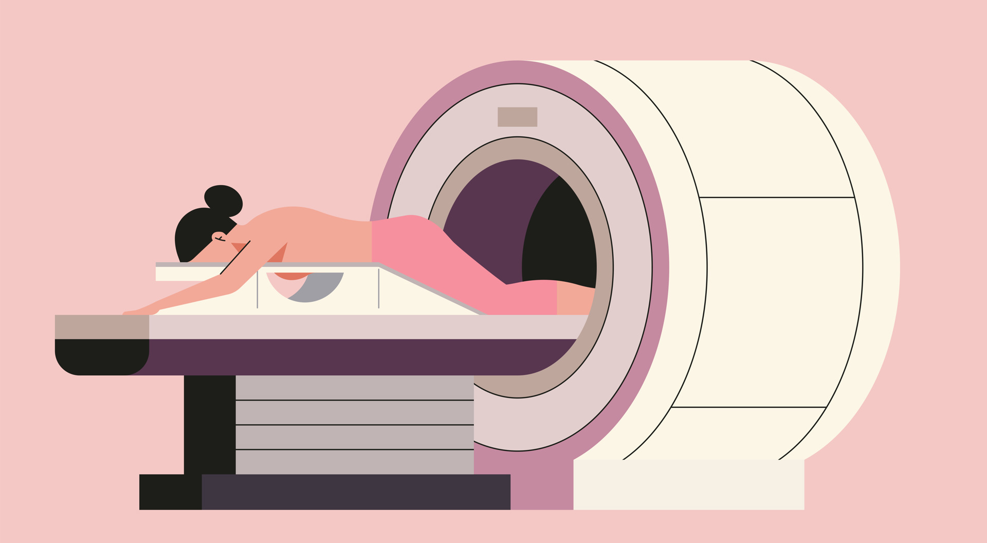
The typical MRI machine for breast imaging is a large, cylindrical tube enclosed by a circular magnet. You'll lie face down on a specialized table with openings for your breasts, which then slides into the tunnel-like opening of the machine. This positions you comfortably within the magnetic field for optimal imaging.
However, there are variations in MRI machine designs to accommodate different patient needs and preferences. Some MRI units, referred to as short-bore systems, offer a more open configuration where the magnet doesn't fully encircle you. This can be less confining and potentially more comfortable for some individuals.
Additionally, newer MRI machines often feature larger diameter bores, providing increased space and comfort for larger patients or those who experience claustrophobia. "Open" MRI units take this concept further by having open sides, creating a significantly less enclosed environment. These open MRI units are particularly beneficial for individuals with anxiety related to enclosed spaces or those with larger body sizes.
While open MRI machines can produce high-quality images for many types of breast exams, their applicability might be limited for certain specialized procedures. To determine the most suitable MRI unit for your specific needs, consulting with your radiologist is recommended. They can provide personalized guidance and address any questions or concerns you may have about the equipment.
How does the procedure work?
Unlike X-rays and computed tomography (CT) scans, which utilize ionizing radiation, MRI employs a powerful magnetic field and radio waves to create detailed images of the body's internal structures. This non-invasive technique involves temporarily realigning the hydrogen atoms naturally present within the body's tissues. As these atoms return to their normal state, they release varying amounts of energy, depending on the type of tissue they are in. This energy is captured by the scanner and used to create a detailed picture.
The MRI machine is equipped with coils that transmit and receive radio waves, which interact with the hydrogen atoms in the body. The magnetic field is generated by passing an electric current through wire coils within the machine, and additional coils may be placed around the specific body part being imaged. It's important to note that the electric current itself does not come into direct contact with the patient.
A sophisticated computer then processes the signals emitted by the hydrogen atoms, creating a series of cross-sectional images, each depicting a thin slice of the body. These images can be viewed from different angles, allowing the radiologist to examine the internal structures in detail. MRI's exceptional ability to differentiate between healthy and diseased tissue makes it a valuable diagnostic tool, often surpassing the capabilities of X-ray, CT, and ultrasound in certain situations.
How is the procedure performed?
Breast MRI exams are typically conducted on an outpatient basis, and you will be positioned face down on a specialized platform with openings for your breasts. This platform is designed to capture images without compressing your breasts and contains the electronics necessary for imaging. Remaining still throughout the exam is crucial, and ensuring comfort can help you relax and avoid unnecessary muscle tension. If you experience any discomfort, communicate it to the technologist to minimize movement during the scan.
Contrast material is not used if the MRI is solely for checking implant rupture. However, for all other purposes, intravenous injection of a contrast agent is necessary for accurate breast cancer detection. In such cases, a doctor, nurse, or technologist will insert an intravenous catheter (IV) into your hand or arm for contrast administration.
Once positioned within the MRI machine, the technologist will operate the scanner from a separate room, but you'll be able to communicate with them through an intercom. If contrast is used, it will be injected after initial scans, and additional images will be taken during or after the injection.
After the exam, you may be asked to wait briefly while the radiologist reviews the images to ensure they are satisfactory. If an IV was used, it will be removed, and a small dressing applied. The imaging session itself takes 30 minutes to an hour, with the entire exam typically lasting around an hour and a half.
In some cases, your doctor might perform MR spectroscopy during the exam, which provides additional information about the chemicals within your body's cells. This can add approximately 15 minutes to the overall exam duration.
What will I experience during and after the procedure?
While breast MRI exams are generally painless, some individuals may find it challenging to remain still for extended periods. Additionally, the enclosed space within the MRI machine can trigger feelings of claustrophobia in certain patients. The scanner itself generates noise, but earplugs or headphones are provided to minimize discomfort.
It's normal for the imaged area to feel slightly warm due to the radiofrequency pulses, and if this becomes bothersome, informing the technologist is recommended. Maintaining stillness is crucial for obtaining clear images, and you'll know when images are being recorded due to the tapping or thumping sounds from the activated coils. You may be asked to hold your breath during specific sequences for optimal image quality. Although you may be able to relax between sequences, maintaining your position is essential.
Despite being alone in the exam room, you'll be under constant observation and in communication with the technologist through a two-way intercom. A "squeeze-ball" is provided for immediate assistance if needed. Some facilities allow a screened companion to stay with you during the scan for added comfort.
The technologist may offer earplugs to reduce the scanner's noise, which includes loud thumping and humming sounds. MRI scanners are well-lit and air-conditioned for your comfort, and some even offer music to help you relax during the exam.
If contrast material is used, a brief sensation of coolness and flushing is normal upon injection. The IV needle might cause slight discomfort, and some bruising might occur afterward. There's a small chance of skin irritation at the IV insertion site.
In most cases, if you haven't been sedated, there's no recovery period needed, and you can resume your normal activities and diet immediately. Side effects from the contrast material are rare, but some individuals might experience nausea or local pain. Allergic reactions like hives or itchy eyes are exceedingly uncommon, but if you experience any symptoms, promptly alert the technologist for immediate medical assistance.
Who interprets the results and how do I get them?
Following your breast MRI, a radiologist, a physician specializing in interpreting medical images, will meticulously analyze the scans. They will then prepare a comprehensive report detailing their findings and conclusions, which will be sent to the doctor who referred you for the MRI or your primary care physician. Your doctor will discuss the results with you in detail, explaining any identified abnormalities or concerns, and outline the next steps in your care, whether it involves further testing or treatment.
In some cases, a follow-up MRI or additional imaging tests may be recommended. This could be to further investigate a suspicious finding, monitor any changes over time, or evaluate the effectiveness of an ongoing treatment plan. Follow-up exams are often crucial for ensuring a precise diagnosis and determining the most appropriate course of action for your breast health.
Benefits
Breast MRI offers several key benefits as a diagnostic tool. Notably, it is a non-invasive procedure that does not involve exposure to ionizing radiation, making it a safe option for repeated imaging. MRI has proven invaluable in detecting and staging breast cancer, especially when other imaging techniques like mammography and ultrasound fail to provide sufficient information. This makes it a particularly useful tool for women at high risk of breast cancer, as it can identify early-stage cancers that might be missed by other methods.
MRI excels at imaging dense breast tissue, which is common in younger women and can make mammograms more difficult to interpret. Additionally, MRI can effectively image breast implants, allowing for the detection of ruptures or other complications. In cases where a suspicious lesion is only visible on MRI, the technology can also guide biopsy procedures for accurate diagnosis.
Furthermore, the gadolinium-based contrast material used in some MRI scans is less likely to cause allergic reactions than the iodine-based contrasts used in X-rays and CT scans, making it a safer option for patients with known allergies or sensitivities.
Risks
Breast MRI exams are generally safe for most individuals when appropriate safety guidelines are followed. However, it's important to be aware of potential risks. While the use of sedation carries a small risk of over-sedation, vigilant monitoring of vital signs helps minimize this concern.
The strong magnetic field used in MRI poses no direct harm, but it can potentially interfere with the function of implanted medical devices or distort the resulting images. A rare complication associated with gadolinium-based contrast agents is nephrogenic systemic fibrosis, primarily observed in patients with pre-existing kidney issues. To mitigate this risk, doctors carefully assess kidney function before considering a contrast injection.
Allergic reactions to the contrast material are also possible, although they are typically mild and can be managed with medication. If you experience an allergic reaction, medical professionals will be readily available to provide immediate assistance. Recent research suggests that trace amounts of gadolinium might remain in the body, particularly in the brain, after multiple MRI scans. This is more likely to happen in patients who undergo frequent MRI exams over their lifetime for monitoring chronic or high-risk health conditions. Gadolinium is mainly excreted through the kidneys, but if you fall into this category, discuss the possibility of gadolinium retention with your doctor, as the effects can vary from person to person.
For breastfeeding mothers, manufacturers of IV contrast advise a 24-48 hour pause in breastfeeding after contrast administration. However, the latest guidance from the American College of Radiology indicates that the amount of contrast absorbed by infants through breast milk is minimal. For detailed information and guidance, consult the ACR Manual on Contrast Media and its references.
What are the limitations of a Breast MRI?
While breast MRI offers significant advantages in breast imaging, it is not without limitations. The quality of the images obtained is highly dependent on your ability to remain perfectly still and follow breath-holding instructions during the scan. If you experience anxiety, confusion, or severe pain, maintaining stillness can be difficult, potentially compromising image quality.
Additionally, body size can be a limiting factor, as some MRI machines have weight restrictions, making it challenging for larger individuals to undergo the scan. Implants or other metallic objects in the body can also interfere with image quality, as can patient movement during the procedure. In some cases, an irregular heartbeat might affect the quality of images due to the reliance of some MRI techniques on the heart's electrical activity for timing.
While non-contrast MRI is generally considered safe for pregnant women, doctors may choose to postpone the exam until after delivery if it's not urgent. Gadolinium contrast agents are typically avoided during pregnancy except in specific circumstances. Your doctor will discuss the potential benefits and risks of any MRI procedure with you. MRI might be performed after the first trimester to assess the fetus for findings not fully evaluated by ultrasound.
One notable limitation of breast MRI is its occasional difficulty in distinguishing between cancerous tissue and benign fluid accumulation known as edema. This can sometimes lead to false-positive results, requiring further testing or biopsy to confirm the diagnosis. Furthermore, MRI exams are typically more expensive and time-consuming than other imaging modalities like mammography or ultrasound. If you have concerns about the cost, it's advisable to discuss them with your insurance provider.