Overview of Abdomen/Pelvis MRI
Abdominal and pelvic MRI, a non-invasive imaging technique, utilizes a powerful magnetic field and radio waves to create detailed visualizations of the internal organs and tissues within these regions. This versatile tool aids in the diagnosis and monitoring of a wide spectrum of conditions, from tumors and infections to inflammatory diseases and vascular abnormalities. The procedure is notably safe for pregnant women, offering a radiation-free alternative for monitoring maternal and fetal health during pregnancy. By providing high-resolution images, MRI enables physicians to assess organ structures, detect subtle lesions, and evaluate blood flow patterns, ultimately leading to accurate diagnoses and informed treatment decisions for patients with abdominal or pelvic concerns.
What are some common uses of the procedure?
Abdominal and pelvic MRI serves as a versatile diagnostic tool, offering a comprehensive view of the intricate structures within these body regions. It's employed to assess a wide range of organs, including the liver, biliary tract, kidneys, spleen, pancreas, adrenal glands, bladder, and reproductive organs (uterus and ovaries in females, prostate gland in males). In addition, it can provide valuable insights into the health of blood vessels and lymph nodes, which are critical components of the circulatory and immune systems.
MRI's applications in the abdomen and pelvis extend to diagnosing and monitoring various conditions. In the digestive system, it aids in identifying and staging tumors, evaluating the extent of liver diseases like cirrhosis, and pinpointing abnormalities in the pancreas and bile ducts. It is also instrumental in assessing inflammatory bowel diseases, such as Crohn's disease and ulcerative colitis, providing detailed information about the affected areas.
Moreover, MRI's unique ability to visualize blood vessels non-invasively allows for the detection of abnormal blood vessels, inflamed vessels (vasculitis), and other vascular conditions like aneurysms. In the realm of women's health, it plays a crucial role in monitoring fetal development during pregnancy and diagnosing a wide range of gynecological conditions, including uterine fibroids and pelvic masses. In men, it aids in the diagnosis and staging of prostate cancer.
Overall, abdominal and pelvic MRI is an indispensable tool in modern medicine, offering a comprehensive and non-invasive approach to diagnosing and managing a wide array of conditions within these complex regions of the body.
How should I prepare for the Abdomen/Pelvis MRI?
Preparing for an abdominal and pelvic MRI involves several important steps to ensure a safe and successful procedure. You'll be asked to change into a hospital gown to avoid any image artifacts and comply with safety regulations concerning the strong magnetic field. While you can usually continue your regular medication and diet, guidelines regarding eating and drinking before the exam might vary, so it's best to follow your doctor's or the facility's specific instructions.
In some cases, an injection of gadolinium contrast material may be used to enhance image clarity. If your exam requires contrast, you'll be asked about any allergies or health conditions like asthma, as well as allergies to medications, food, or environmental factors. Gadolinium is generally safe with a lower risk of allergic reactions than iodine-based contrast, but your doctor will carefully assess your medical history before administering it.
It's crucial to inform the technologist or radiologist about any serious health issues, such as kidney problems or recent surgeries, as these factors might influence the use of contrast or the overall safety of the procedure. Pregnant women should always inform their doctor and the technologist, as MRI is generally safe but requires special considerations during the first trimester. Gadolinium contrast is typically avoided unless absolutely necessary.
If you experience claustrophobia or anxiety, discuss the possibility of a mild sedative with your doctor before the exam. For infants and young children, sedation or anesthesia may be necessary to ensure they remain still during the scan. The specific requirements depend on the child's age, development, and the type of exam. Pediatric sedation specialists are available at many facilities to ensure their safety and comfort.
To minimize anxiety and the need for sedation in children, some facilities employ certified child life specialists. These professionals, with backgrounds in child development and psychology, can prepare children for the MRI experience through explanations and demonstrations, using model scanners and playing the sounds they might hear during the scan.
Many facilities also offer child-friendly imaging suites with comforting décor and utilize techniques like silent MRI and distraction tools such as DVD goggles or music headphones. Advancements in MRI technology have also led to shorter scan times, which can be beneficial for children.
Before entering the MRI room, remove all jewelry, accessories, and any metal or electronic items. These can interfere with the magnetic field, potentially causing damage or becoming projectiles. This includes items like jewelry, watches, credit cards, hearing aids, hairpins, metal zippers, removable dental work, pens, pocketknives, eyeglasses, body piercings, and electronic devices like mobile phones and smartwatches.
Most metal implants are safe in MRI, but exceptions include certain cochlear implants, aneurysm clips, metal coils in blood vessels, older cardiac devices, and vagal nerve stimulators. Inform the technologist of any implanted devices and provide any relevant documentation. If needed, an X-ray can be used to identify metal objects.
If you have any shrapnel, bullets, or other metal fragments in your body, especially near the eyes, inform the medical staff. These can be hazardous due to potential movement or heating during the scan. While tattoo dyes containing iron might rarely cause issues, most dental fillings, braces, and cosmetics are generally safe, although they may affect image quality in the facial or brain areas. Finally, anyone accompanying you into the MRI room will also be screened for metal and electronic devices to ensure a safe environment.
What does the equipment look like?
The typical cardiac MRI machine is a large, cylindrical tube enclosed by a circular magnet, similar to other MRI units. During the scan, you'll lie on a movable table that slides into the center of this tunnel-like structure.
However, alternative designs exist to accommodate patient comfort and specific needs. Short-bore systems offer a more open configuration where the magnet doesn't completely surround you, providing a less confined experience. Additionally, newer MRI machines often feature larger diameter bores, allowing for more space and comfort, particularly for larger patients or those with claustrophobia.
"Open" MRI units represent a further departure from the traditional design, with open sides that create a significantly less claustrophobic environment. These units are particularly beneficial for individuals who experience anxiety in enclosed spaces or those with larger body sizes. While open MRI machines can produce high-quality images for many cardiac exams, their suitability for specific procedures may vary. It's advisable to consult with your radiologist to determine the most appropriate MRI unit for your individual needs.
How does the procedure work?
Unlike X-rays and computed tomography (CT) scans, which utilize ionizing radiation, MRI employs a powerful magnetic field and radio waves to create detailed images of the body's internal structures. This non-invasive technique works by temporarily realigning the hydrogen atoms naturally present within the body's tissues. As these atoms return to their normal alignment, they release varying amounts of energy, depending on the type of tissue they are in. This energy is detected by the scanner and used to construct a detailed image.
The MRI machine is equipped with coils that transmit and receive radio waves, which interact with the hydrogen atoms in the body. The magnetic field is generated by passing an electric current through wire coils within the machine, and additional coils may be placed around the specific body part being imaged. It's important to note that the electric current itself does not come into direct contact with the patient.
A sophisticated computer then processes the signals emitted by the hydrogen atoms, creating a series of cross-sectional images, each depicting a thin slice of the body. These images can be viewed from different angles, allowing the radiologist to examine the internal structures in detail. MRI's exceptional ability to differentiate between healthy and diseased tissue makes it a valuable diagnostic tool, often surpassing the capabilities of X-ray, CT, and ultrasound in certain situations.
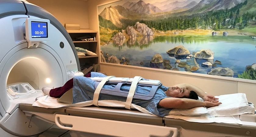
How is the procedure performed?
Abdominal and pelvic MRI exams can be conducted on both outpatients and inpatients. You will be positioned on a movable table by the technologist, who may use straps and bolsters to help you remain still throughout the scan. Specialized coils that send and receive radio waves may be placed around or near your abdomen and pelvis to enhance image quality.
The exam typically involves multiple sequences, each producing distinct sounds and lasting several minutes. If your exam requires contrast material, a doctor, nurse, or technologist will insert an intravenous catheter (IV) into a vein in your hand or arm to administer it. You'll then be placed inside the MRI machine, where the technologist will operate the scanner from a separate room. You'll be able to communicate with them through an intercom throughout the exam.
In some cases, oral contrast may be used to better visualize the bowel during specific MRI exams like MR Enterography. If contrast material is used, whether oral or intravenous, additional images will be taken after its administration.
Upon completion of the scan, you might be asked to wait briefly while the radiologist reviews the images to ensure they are of sufficient quality. The technologist will then remove the IV line, if applicable, and apply a small dressing to the insertion site.
The entire exam duration can vary depending on the type of exam and the equipment used, but it generally takes between 30 to 50 minutes.
What will I experience during and after the procedure?
While abdominal and pelvic MRI exams are generally painless, some individuals may find it challenging to remain still for extended periods. Additionally, the enclosed space within the scanner can trigger feelings of claustrophobia in certain patients. The scanner itself generates noise, but earplugs or headphones are provided to minimize discomfort.
During the scan, a warming sensation in the imaged area is normal due to the radiofrequency pulses, and you can inform the technologist if it becomes uncomfortable. Maintaining stillness is crucial, particularly during the brief image recording periods, which are accompanied by loud tapping or thumping sounds from the activated coils. Although you may be able to relax between sequences, minimizing movement is essential for clear images.
You'll be alone in the exam room but in constant communication with the technologist through a two-way intercom. A "squeeze-ball" is provided for immediate assistance if needed, and some facilities allow a screened companion to stay with you.
For children, appropriately sized earplugs or headphones are provided, and music might be played to help them relax. MRI scanners are well-lit and air-conditioned for comfort.
In some cases, contrast material is injected intravenously (IV) before the scan. This may cause slight discomfort, bruising, or a temporary metallic taste.
If sedation is not required, there's no recovery period, and you can resume your normal activities and diet immediately. Side effects from contrast material are rare but can include nausea, headache, or pain at the injection site. Allergic reactions like hives or itchy eyes are exceedingly rare but require immediate medical attention.
Who interprets the results and how do I get them?
Following your abdominal and pelvic MRI, a radiologist, a doctor specializing in interpreting medical images, will meticulously analyze the scans. They will then compile a detailed report outlining their findings and conclusions, which will be sent to your primary care physician or the doctor who referred you for the MRI. Your doctor will discuss the results with you, explaining any identified abnormalities or issues in detail and outlining any necessary next steps.
In some cases, a follow-up MRI or additional imaging tests may be recommended. This could be to further investigate a suspicious finding, monitor any changes over time, or assess the effectiveness of an ongoing treatment plan. Follow-up exams are often crucial for ensuring accurate diagnosis and effective management of any abdominal or pelvic conditions that may be identified during the initial MRI.
Benefits
Abdominal and pelvic MRI offers several key benefits as a diagnostic tool. Notably, it's a non-invasive procedure that doesn't expose patients to ionizing radiation, making it a safe option for repeated imaging. MRI excels at producing highly detailed images of soft-tissue structures, such as the liver and other organs, often surpassing other imaging methods in accurately identifying and characterizing diseases. This makes it invaluable for early diagnosis and evaluation of various abnormalities, including tumors.
MRI's versatility extends to diagnosing a wide range of conditions, including cancer, heart and vascular diseases, and musculoskeletal disorders. It can detect abnormalities that might be hidden by bone in other imaging techniques, providing a more comprehensive view of the abdominal and pelvic regions. Additionally, MRI allows for non-invasive assessment of the biliary system without the need for contrast injection, further enhancing its safety profile.
The gadolinium-based contrast material used in some MRI scans is less likely to cause allergic reactions than iodine-based contrasts used in X-rays and CT scans, making it a suitable option for patients with known allergies or sensitivities. Finally, MRI offers a non-invasive alternative to X-ray angiography and CT for diagnosing blood vessel problems, reducing the risks associated with more invasive procedures.
Risks
Abdominal and pelvic MRI scans are generally considered safe for most patients when appropriate safety guidelines are followed. However, certain risks should be acknowledged. While the use of sedation carries a small risk of over-sedation, vigilant monitoring of vital signs helps minimize this concern.
The strong magnetic field used in MRI poses no direct harm but can potentially interfere with the function of implanted medical devices or distort the resulting images. A rare complication associated with gadolinium-based contrast agents is nephrogenic systemic fibrosis, primarily observed in patients with pre-existing kidney issues. To mitigate this risk, doctors carefully assess kidney function before considering a contrast injection.
Allergic reactions to the contrast material are also possible, although they are typically mild and can be managed with medication. Medical assistance is readily available in case of an allergic reaction. Recent research suggests that trace amounts of gadolinium might remain in the body, particularly in the brain, after multiple MRI scans. This is more likely to happen in patients who undergo frequent MRI exams over their lifetime for monitoring chronic or high-risk health conditions. Gadolinium is mainly excreted through the kidneys, but if you fall into this category, discuss the possibility of gadolinium retention with your doctor, as the effects can vary from person to person.
For breastfeeding mothers, manufacturers of IV contrast advise a 24-48 hour pause in breastfeeding after contrast administration. However, the latest guidance from the American College of Radiology indicates that the amount of contrast absorbed by infants through breast milk is minimal. For detailed information and guidance, consult the ACR Manual on Contrast Media and its references.
What are the limitations of a Abdomen/Pelvis MRI?
While abdominal and pelvic MRI offers valuable diagnostic insights, it's important to be aware of its limitations. The quality of the images obtained relies heavily on the patient's ability to remain perfectly still and follow breath-holding instructions during the scan. If you experience anxiety, confusion, or severe pain, maintaining stillness can be challenging, potentially compromising image clarity.
Body size can also be a constraint, as some MRI machines have limited openings, making it difficult for larger individuals to undergo the scan. Additionally, the presence of metal implants or other metallic objects in the body can interfere with image quality, as can patient movement during the procedure. Breathing and bowel movements can also cause artifacts or image distortions, although modern scanners and techniques have minimized this issue.
For pregnant women, non-contrast MRI is generally considered safe, with no conclusive evidence of harm to the fetus. However, doctors may opt to postpone the exam until after delivery if it's not urgent. Gadolinium contrast agents are typically avoided during pregnancy except in specific circumstances. Your doctor will discuss the potential benefits and risks of any MRI procedure with you.
Another limitation of MRI is its occasional difficulty in distinguishing between cancerous tissue and benign tumors or other conditions, such as edema. This can sometimes necessitate additional testing or imaging modalities for a definitive diagnosis. Lastly, MRI exams tend to be more expensive and time-consuming than other imaging techniques like ultrasound or X-rays. If cost is a concern, it's advisable to discuss it with your insurance provider.