Overview of X-ray IVP - Intravenous Pyelogram
An Intravenous Pyelogram (IVP), also known as an Intravenous Urogram (IVU), is a specialized X-ray examination that uses a contrast material (dye) injected into a vein to visualize the entire urinary tract. This includes the kidneys, which filter waste from the blood and produce urine; the ureters, tubes that carry urine from the kidneys to the bladder; and the bladder, which stores urine. Once injected, the iodine-based contrast material travels through the bloodstream and is filtered by the kidneys, highlighting these structures on X-ray images as it passes through. A series of X-rays are taken at timed intervals as the contrast flows through the urinary system. This allows radiologists to assess the size, shape, and position of these organs, detect abnormalities, and evaluate the flow of urine. It is a key diagnostic tool for various urinary system disorders.
Why an X-ray IVP is Done
An X-ray IVP is performed to investigate a range of symptoms and conditions affecting the urinary tract. It is commonly ordered to diagnose the cause of blood in the urine (hematuria), flank or lower abdominal pain, or recurrent urinary tract infections. Conditions it helps detect include kidney stones or stones in the ureters or bladder, blockages (obstructions) in the urinary tract, kidney cysts, tumors in the kidneys or bladder, and structural abnormalities of the urinary system (such as congenital anomalies). It can also assess for kidney injury following trauma or help locate the source of a urinary tract infection. By observing how the contrast material moves through the system, radiologists can identify issues with urine flow, assess the filtering capacity of the kidneys, and pinpoint anatomical problems that might be contributing to a patient's symptoms.
Risks
An X-ray IVP involves exposure to ionizing radiation and the administration of an iodine-based contrast material. The radiation dose is generally low, but there's a small theoretical risk of cell damage. For pregnant women or those who suspect they might be pregnant, it is crucial to inform the doctor and technologist, as the procedure is generally avoided during pregnancy due to potential risks to the fetus. The primary risks are associated with the contrast material. Allergic reactions, ranging from mild (itching, hives, nausea) to severe (difficulty breathing, swelling, anaphylaxis), can occur, though severe reactions are rare. There is also a risk of contrast-induced nephropathy (kidney damage), particularly in patients with pre-existing kidney disease, diabetes, or dehydration. Patients with a history of allergies, asthma, or kidney problems should inform their doctor, as precautions or alternative imaging might be necessary.
How You Prepare
Proper preparation is crucial for an accurate X-ray IVP. You will typically be instructed to fast for at least 8 to 12 hours before the procedure, meaning no food or drinks (including water) during this period. This helps ensure that the digestive tract is clear and does not obscure the view of the urinary system. You may also be given a bowel preparation kit (laxatives) to take the evening before the exam to empty your bowels, further improving visibility. It is vital to inform your doctor about all medications you are taking, especially metformin (Glucophage), as it may need to be stopped temporarily. Also, disclose any allergies, particularly to iodine or contrast dyes, and any history of kidney disease, diabetes, or asthma. Most importantly, pregnant women or those who suspect pregnancy must inform their doctor, as the procedure is usually avoided due to radiation exposure to the fetus.
What You Can Expect
Before the Test
Before your X-ray IVP, you will receive detailed preparation instructions, typically including fasting for 8 to 12 hours and possibly taking a bowel preparation the evening prior to clear your digestive tract. You will be asked to remove any metal objects, such as jewelry, eyeglasses, or dentures, and may be provided with a hospital gown. It is crucial to inform the technologist about any allergies (especially to iodine or contrast dye), existing medical conditions (like diabetes or kidney disease), and most importantly, if you are pregnant or suspect you might be. The technologist will explain the procedure, answer your questions, and may measure your blood pressure and heart rate. You might also have a blood test to check your kidney function before the contrast is administered, ensuring it's safe to proceed.
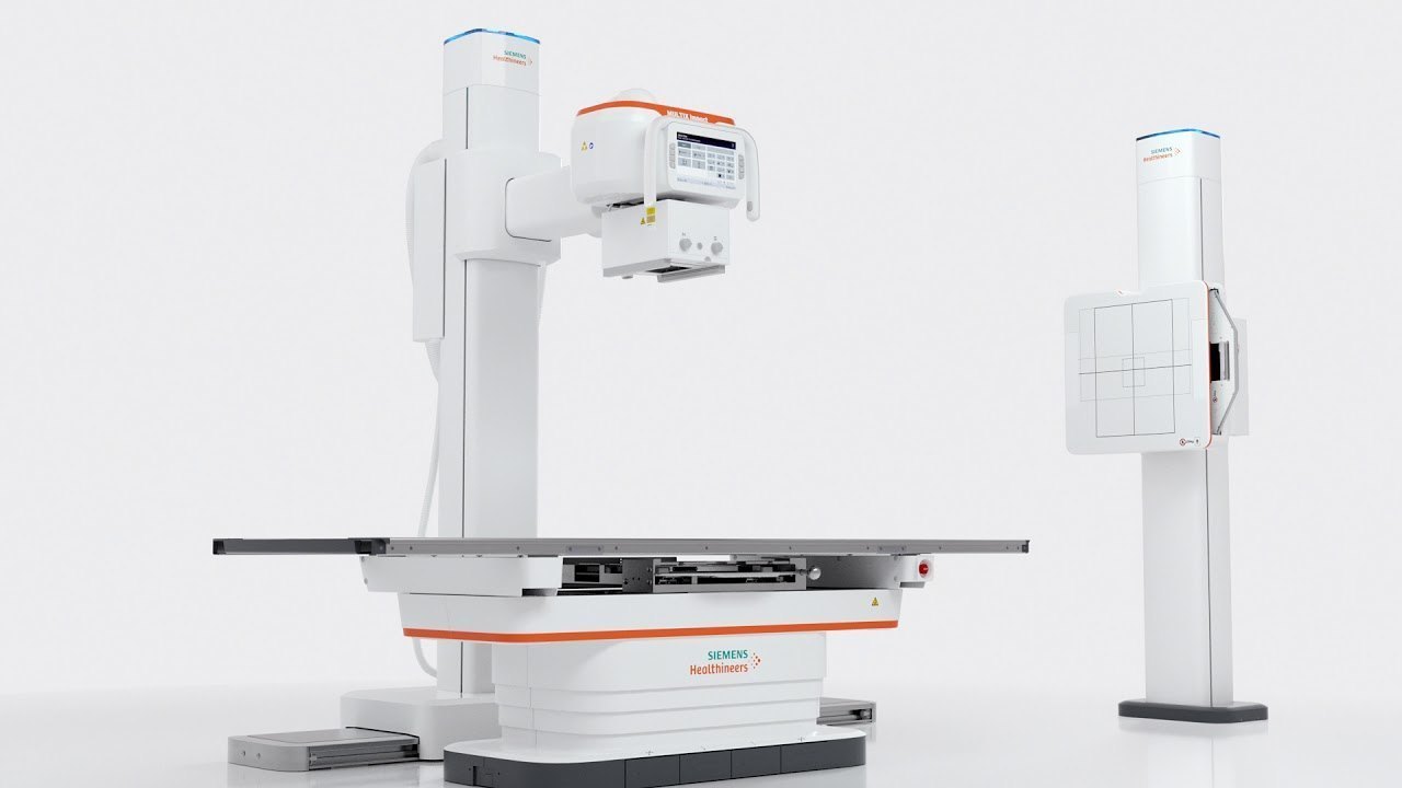
During the Test
During an X-ray IVP, you will lie on an X-ray table. A trained technologist will insert an intravenous (IV) line into a vein, usually in your arm. The iodine-based contrast material will then be injected through this IV line. You may feel a warm flush sensation throughout your body, a metallic taste in your mouth, or a temporary feeling of nausea as the contrast is injected; these are normal sensations. A series of X-ray images will be taken at timed intervals as the contrast flows through your kidneys, ureters, and bladder. The technologist may gently apply a compression band over your abdomen (unless contraindicated) to help the kidneys retain the contrast for better visualization. You may also be asked to change positions, take deep breaths, or hold your breath briefly. The entire procedure typically takes about 30 to 60 minutes, though it can sometimes extend longer for delayed images.
Results
After your X-ray IVP is completed, the IV line will be removed, and the images will be sent to a radiologist, a medical doctor specially trained in interpreting X-ray and other imaging studies. The radiologist will carefully analyze the series of images, observing how the contrast material filled and passed through your kidneys, ureters, and bladder. They will assess the size, shape, and position of these organs, looking for any abnormalities such as kidney stones, blockages, narrowing (strictures), cysts, tumors, or anatomical variations. A detailed report of these findings will then be prepared and sent to your referring healthcare provider, usually within a few days. Your doctor will then discuss the results with you, explain what they mean for your urinary symptoms or condition, and recommend any necessary further tests, treatments, or management plans based on the IVP findings. You will be advised to drink plenty of fluids after the test to help flush the contrast material from your system.