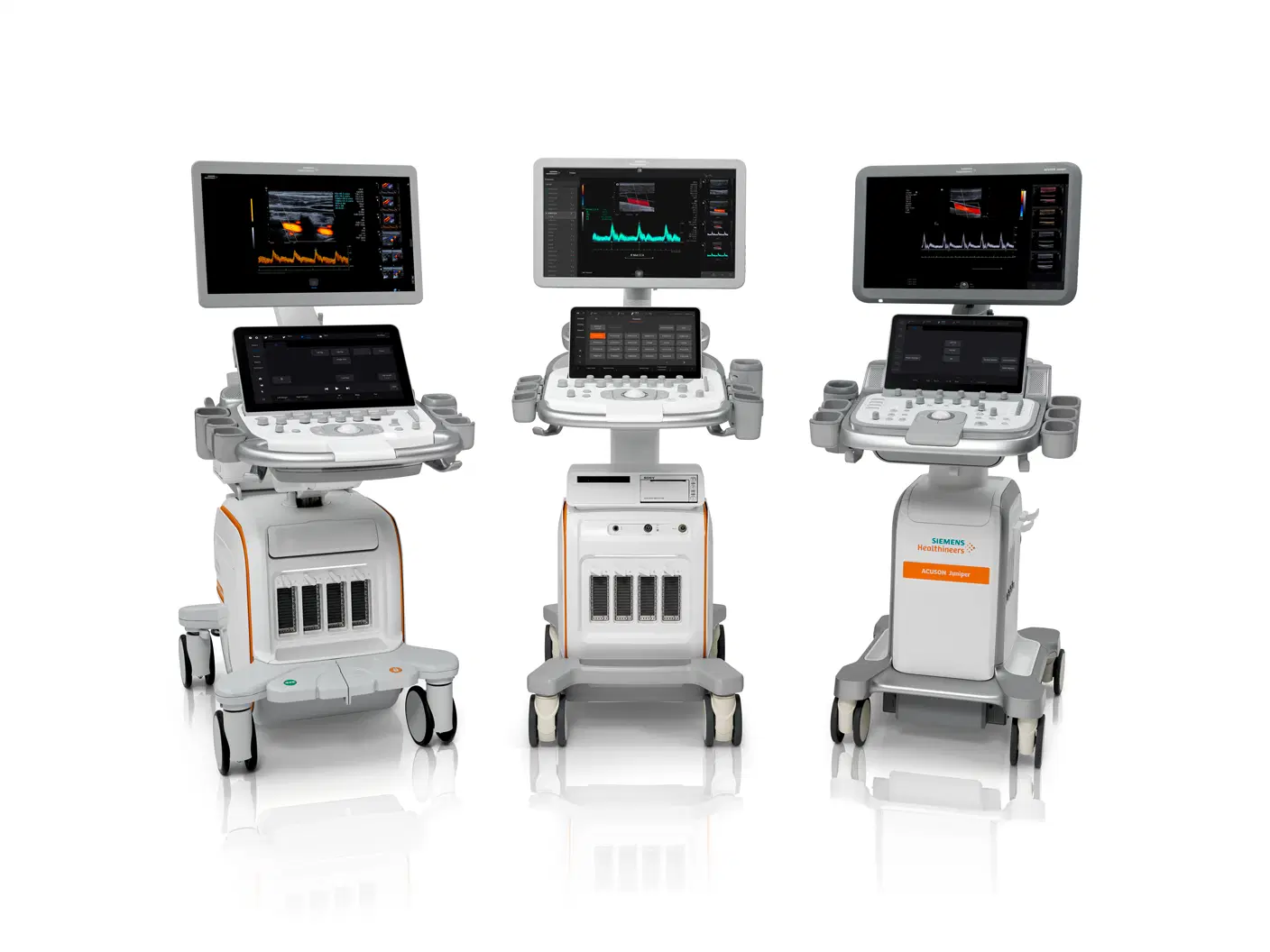
Overview of Sonography
Diagnostic ultrasound, also known as sonography, is a versatile and non-invasive medical imaging technique that uses high-frequency sound waves to visualize the internal structures of the body. These sound waves, emitted by a transducer, bounce off tissues and organs, creating echoes that are then processed by a computer to generate real-time images or videos. This valuable tool aids healthcare professionals in diagnosing a wide range of conditions, from monitoring the health of a developing fetus during pregnancy to identifying tumors, evaluating blood flow, and guiding biopsies.
Ultrasound exams are typically performed with a transducer placed on the skin's surface, often with a lubricating gel to enhance sound transmission. However, in some cases, a small probe may be inserted into a body cavity for specialized imaging, such as transvaginal or transrectal ultrasound. The safety and painless nature of ultrasound make it a preferred imaging modality for various medical applications, offering valuable insights into the body's inner workings without the need for radiation exposure or invasive procedures.
Why it's Done
Ultrasound's versatility makes it a cornerstone of prenatal care, allowing expectant mothers to witness the miracle of life unfolding within them. By visualizing the uterus and ovaries, healthcare providers can monitor fetal development throughout pregnancy, ensuring the baby's well-being. Ultrasound also plays a vital role in diagnosing a multitude of conditions. It can detect gallstones and other abnormalities within the gallbladder, assess blood flow to determine potential blockages or vascular problems, and guide needles with exceptional precision during biopsies or tumor treatments. This non-invasive technique is also employed for examining breast lumps, a crucial step in early cancer detection. It can be used to evaluate the health of the thyroid gland, a butterfly-shaped organ located at the base of the neck that produces hormones regulating metabolism and growth. In the realm of urology, ultrasound aids in diagnosing issues with the genitals and prostate, helping identify potential problems early on. Furthermore, ultrasound proves its worth in assessing joint inflammation, a condition known as synovitis, and evaluating metabolic bone diseases that affect bone mineral density and structure. By offering a safe and effective way to visualize internal structures and guide medical procedures, ultrasound remains an indispensable tool in modern medicine.
Risks
Diagnostic ultrasound boasts an impressive safety record, employing low-power sound waves that pose no known risks to patients. Its non-invasive nature and absence of radiation exposure make it a preferred choice for various medical applications, particularly during pregnancy and for sensitive populations like children and individuals with weakened immune systems. However, it's important to acknowledge that ultrasound, like any medical tool, has its limitations.
The very nature of sound waves, which are the foundation of ultrasound imaging, poses challenges when encountering air or bone. These substances act as barriers, impeding the transmission of sound waves and hindering their ability to generate clear images. For instance, gas-filled organs like the lungs can significantly distort the sound waves, making it difficult to visualize underlying structures. Similarly, sound waves struggle to penetrate bone effectively, limiting their ability to image organs and tissues encased within the bony structures of the skull or pelvis.
Additionally, ultrasound's reach may be restricted when attempting to visualize structures located deep within the body. Sound waves tend to lose energy as they travel through tissue, and their ability to penetrate diminishes with increasing depth. This can make it challenging to obtain detailed images of organs situated deep within the abdomen or pelvis. In such cases, healthcare professionals may opt for complementary imaging modalities like CT scans, MRI scans, or X-rays, each offering unique strengths in visualizing different tissues and organs. CT scans, for example, excel at capturing detailed cross-sectional images of the body, while MRI scans provide unparalleled views of soft tissues and organs. X-rays, on the other hand, are particularly effective in imaging bones and detecting fractures.
How you Prepare
In most cases, preparing for an ultrasound is as simple as showing up for your appointment. However, there are a few scenarios where specific instructions might apply. If you're scheduled for a gallbladder ultrasound, your doctor may advise you to abstain from food and drink for a certain period beforehand to ensure optimal image quality. On the other hand, some procedures, like pelvic ultrasounds, may require a full bladder to provide better visualization of the organs. In this instance, your healthcare provider will guide you on how much water to drink and when to start drinking it.
For younger patients, additional preparation measures may be necessary. It's always best to consult your healthcare provider when scheduling an ultrasound for yourself or your child to receive tailored instructions. On the day of your appointment, choose comfortable, loose-fitting clothing that allows easy access to the area being examined. Be prepared to remove any jewelry in the vicinity of the scan, and it's wise to leave valuables at home for added convenience. By following these guidelines, you can ensure a smooth and efficient ultrasound experience.
What you can Expect
Before the Test
Upon arrival for your ultrasound appointment, you'll be greeted by a friendly healthcare professional who will guide you through the process. In preparation for the scan, you may be asked to remove any jewelry that might interfere with the imaging process and either remove or adjust your clothing to expose the area to be examined. A gown may be provided for your comfort and privacy. Once you're ready, you'll be invited to lie down on a comfortable examination table, where the sonographer will position you optimally for the procedure.
During the Test
The ultrasound procedure begins with the application of a clear, water-based gel to the skin over the area being examined. This gel acts as a conduit, ensuring smooth transmission of the sound waves and preventing air pockets from interfering with image quality. It's safe, easily removed, and won't stain your clothing. A skilled sonographer then employs a handheld device called a transducer, gently pressing it against your skin and maneuvering it to capture images from various angles. The transducer emits high-frequency sound waves that penetrate the body, bouncing off internal structures and returning as echoes. These echoes are then converted into real-time images displayed on a monitor, allowing the sonographer to assess the targeted area.
In some cases, specialized ultrasound exams may require internal imaging. For instance, a transesophageal echocardiogram utilizes a transducer inserted into the esophagus to obtain detailed images of the heart. This procedure is typically performed under sedation to ensure patient comfort. Transrectal ultrasound involves placing a specialized transducer into the rectum to visualize the prostate, while transvaginal ultrasound utilizes a transducer inserted into the vagina to assess the uterus and ovaries.
While most ultrasound exams are painless, you might experience mild discomfort as the sonographer guides the transducer over your body. A full bladder or internal ultrasound probes can sometimes cause slight discomfort, but it's generally temporary and well-tolerated. The duration of a typical ultrasound exam can range from 30 minutes to an hour, depending on the complexity of the examination and the number of areas being assessed.
Results
Following the ultrasound examination, the captured images are meticulously analyzed by a radiologist, a physician specializing in interpreting diagnostic imaging studies. The radiologist then prepares a detailed report, which is sent to your referring healthcare provider. Your doctor will discuss the results with you, explaining any findings or necessary follow-up steps. In most cases, you can resume your normal activities immediately after the ultrasound, as there is no recovery period required. The non-invasive nature of this procedure ensures minimal disruption to your daily routine.