Overview of Brain MRI
Magnetic Resonance Imaging (MRI), particularly of the brain or head, is a sophisticated medical imaging technique renowned for its ability to generate remarkably clear and detailed pictures of the brain's intricate structures. Unlike traditional X-rays or CT scans, MRI harnesses the power of a strong magnetic field, radiofrequency pulses, and a computer to create these images without exposing the patient to ionizing radiation. This makes it a safe and preferred option for diagnosing a wide range of brain disorders, from tumors and strokes to infections and developmental anomalies.
The exceptional clarity and detail provided by MRI scans enable doctors to delve deeper into the complexities of the brain, identifying subtle changes that might be missed by other imaging modalities. This level of precision is crucial for accurate diagnosis and treatment planning, particularly for neurological conditions where early detection can significantly impact patient outcomes. In some cases, a contrast agent called gadolinium may be injected to enhance the visibility of specific tissues or abnormalities, further aiding in the diagnostic process. With its non-invasive nature and remarkable imaging capabilities, MRI has become an indispensable tool in the field of neurology, offering invaluable insights into the inner workings of the human brain.
Why it's done
Brain MRI is a powerful diagnostic tool employed by doctors to investigate a wide array of neurological conditions, whether they manifest suddenly or have been present for an extended period. Its ability to visualize the brain's intricate structures with exceptional clarity makes it invaluable in diagnosing stroke and assessing the extent of stroke-related damage. Additionally, it can detect subtle changes associated with aging, such as brain volume loss or alterations in brain tissue signals, shedding light on the aging process and its impact on cognitive function.
In cases of traumatic brain injury, MRI helps identify the location and severity of damage, including bleeding and bruising, aiding in the development of appropriate treatment plans. For individuals experiencing a loss of body movement control, a condition known as ataxia, MRI can pinpoint the underlying cause, whether it's a lesion, tumor, or other neurological abnormality. Furthermore, brain MRI is instrumental in detecting and characterizing brain tumors, determining their size, location, and potential for malignancy. It also plays a crucial role in diagnosing developmental anomalies that may have arisen during fetal development and identifying hydrocephalus, a condition characterized by the accumulation of fluid within the brain's ventricles.
For those suffering from epilepsy or seizures, MRI can uncover the underlying causes, such as scar tissue, malformations, or tumors, facilitating the selection of appropriate treatment strategies. It also aids in diagnosing brain infections, pinpointing the location and extent of inflammation, and guiding treatment decisions. For chronic conditions like multiple sclerosis, MRI allows for monitoring disease progression and assessing the effectiveness of therapeutic interventions. Beyond the brain itself, MRI can also reveal abnormalities in the eyes and inner ear, providing valuable diagnostic information for a range of ophthalmological and otological disorders. Moreover, it plays a pivotal role in evaluating the pituitary gland, a small but crucial endocrine organ nestled at the base of the brain that regulates hormone production.
How should I prepare for the brain MRI?
Preparing for an MRI scan involves a few key steps to ensure a smooth and safe experience. Firstly, you'll be asked to change into a hospital gown. This is essential to prevent any clothing materials that could interfere with the magnetic field or potentially heat up during the scan from entering the MRI machine.
Secondly, guidelines regarding eating and drinking before the exam can vary, so it's crucial to inquire with your imaging center for specific instructions. In most cases, you can continue taking your medications as usual unless otherwise advised by your doctor. Some MRI scans require an intravenous injection of a contrast material, typically gadolinium, to enhance image clarity. The imaging center will inquire about any allergies you may have to contrast materials to ensure your safety. It's worth noting that gadolinium is generally considered safe, even for individuals with iodine allergies.
Before the scan, inform the technologist of any significant health issues, such as kidney disease or recent surgeries, to assess potential risks and adjust the procedure accordingly. Pregnant women should always inform their doctor and technologist as MRI is generally safe during pregnancy, but certain precautions may be necessary, especially in the first trimester. Additionally, the use of gadolinium contrast in pregnant women is usually limited to essential cases.
If you experience claustrophobia or anxiety, consider discussing with your doctor the possibility of a mild sedative to alleviate any discomfort during the scan. Leaving jewelry and other accessories at home or removing them before entering the MRI room is crucial. Metal and electronic items can disrupt the magnetic field, potentially causing burns or becoming dangerous projectiles. This includes items like jewelry, watches, credit cards, hearing aids, hairpins, metal zippers, removable dental work, pens, pocketknives, eyeglasses, body piercings, magnetic false eyelashes, and electronic devices such as mobile phones and smartwatches.
In most cases, patients with metal implants can safely undergo MRI scans, but exceptions exist. Individuals with certain cochlear implants, aneurysm clips, older cardiac devices, nerve stimulators, or other implanted medical or electronic devices should inform the technologist. In these instances, providing documentation about the implant and its MRI compatibility is essential to assess potential risks and determine the safest course of action. If necessary, an X-ray can be performed to detect and identify any metal objects.
It's also crucial to inform the technologist or radiologist about any shrapnel, bullets, or other metal fragments that may be present in your body, especially near the eyes, as these can move or heat up during the scan, causing harm. While tattoo dyes containing iron might rarely heat up, the magnetic field generally doesn't affect tooth fillings, braces, eyeshadows, or other cosmetics, although they may distort images of the facial area or brain. Therefore, it's important to mention them to the radiologist. Lastly, anyone accompanying you into the exam room must also undergo screening for metal objects and implanted devices to ensure a safe environment for all.
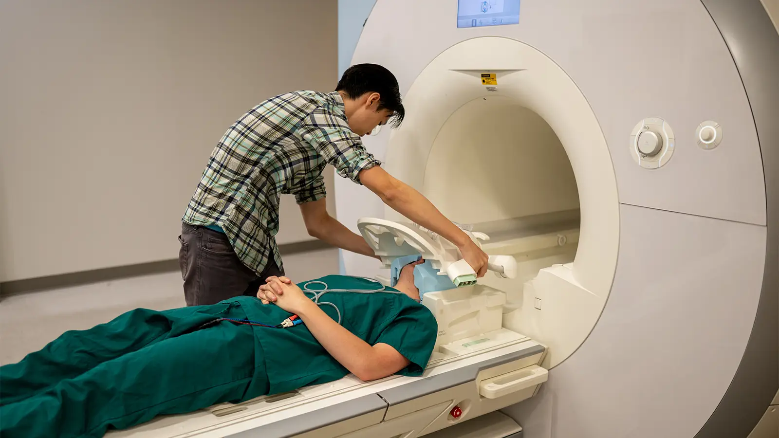
What does the equipment look like?
The classic image of an MRI machine is a large, cylindrical tube enclosed by a circular magnet. During the scan, you lie on a table that smoothly glides into the tunnel-like opening, positioning you within the magnetic field for optimal imaging. However, not all MRI units conform to this traditional design. Some, known as short-bore systems, offer a more open configuration where the magnet doesn't fully encircle you, providing a less confining experience.
Advancements in technology have led to the development of MRI machines with larger diameter bores, accommodating individuals of varying sizes and those who may experience claustrophobia. These wider bores alleviate feelings of confinement and offer a more comfortable experience for many patients. Additionally, "open" MRI units are designed with open sides, providing a significantly less claustrophobic environment, especially beneficial for larger patients or those with anxiety related to enclosed spaces. While open MRI machines can produce high-quality images for numerous examinations, it's important to note that they may not be suitable for all types of scans. If you have concerns about the type of MRI unit best suited for your needs, it's advisable to consult with your radiologist for personalized guidance.
How does the procedure work?
Unlike X-ray and CT scans, MRI doesn't rely on radiation. Instead, it utilizes a powerful magnetic field to temporarily realign hydrogen atoms naturally found in the body. By sending short bursts of radiofrequency energy and measuring the returning signals, a computer generates detailed cross-sectional images of internal structures. The MRI magnet is always on, emphasizing the importance of adhering to safety guidelines. MRI excels at differentiating between healthy and diseased tissue, making it a valuable diagnostic tool.
How is the procedure performed?
Unlike X-ray and CT scans, MRI uses a strong magnetic field and radiofrequency pulses to create detailed images, eliminating the need for radiation. During the scan, you lie within the MRI machine while a technologist operates it from a nearby computer. Various sounds, like clicking or banging, are normal during the imaging process. The machine's powerful magnet aligns hydrogen atoms in your body, and radio waves generate signals that are then converted into images. MRI excels at differentiating between healthy and diseased tissue, making it a valuable diagnostic tool for numerous conditions.
What will I experience during and after the procedure?
While most MRI exams are painless, remaining still for an extended period can be a challenge for some individuals. Additionally, the enclosed space within the scanner might induce claustrophobia in certain patients. The loud noises emitted by the machine are a common experience, but earplugs are provided to minimize discomfort. It's normal for the imaged area to feel slightly warm due to the radiofrequency pulses, and if this becomes bothersome, informing the technologist is recommended. Maintaining stillness during image acquisition is crucial for obtaining clear and accurate results.
Throughout the exam, you'll be in constant communication with the technologist through a two-way intercom, ensuring your safety and addressing any concerns you may have. A "squeeze-ball" is provided as an emergency alert system, offering you immediate access to assistance if needed. Some facilities allow a screened companion to stay with you during the scan, providing an additional layer of comfort. For children, appropriately sized earplugs or headphones are provided, and music may be played to help them relax and pass the time. MRI scanners are well-lit and air-conditioned to ensure a comfortable environment.
In certain cases, an intravenous (IV) injection of contrast material may be administered before the scan to enhance image clarity. While the IV insertion may cause mild discomfort or bruising, the risk of skin irritation is minimal. Some patients may experience a temporary metallic taste after the contrast injection. After the scan, if you haven't received sedation, you can typically resume your regular activities and diet immediately. Although rare, some individuals may experience side effects from the contrast material, including nausea, headache, or pain at the injection site. Allergic reactions like hives or itchy eyes are exceedingly rare, but if you notice any symptoms, promptly alert the technologist, and a radiologist or doctor will be available to provide immediate assistance.
Who interprets the results and how do I get them?
A radiologist, a physician specializing in the interpretation of medical images, meticulously analyzes the MRI scans. Following their comprehensive assessment, a detailed report is sent to the healthcare provider who ordered the exam. Your doctor will then discuss the results with you, elucidating any findings and outlining any necessary follow-up steps. In some instances, a follow-up exam may be recommended to gain a more comprehensive understanding of a potential issue, monitor its progression, or evaluate the effectiveness of ongoing treatment. These follow-up scans often involve additional views or specialized imaging techniques to provide a clearer picture of the situation and guide further decision-making.
Benefits
Magnetic Resonance Imaging (MRI) stands out as a non-invasive diagnostic tool, offering a radiation-free alternative to traditional imaging methods. Its ability to generate exceptionally clear and detailed images of the brain's intricate structures makes it a valuable asset in the medical field. MRI not only allows physicians to assess the brain's anatomy but can also provide functional insights in specific cases, offering a comprehensive understanding of brain activity.
Compared to other imaging modalities, MRI's superior resolution and detail make it an indispensable tool for early disease detection and evaluation, particularly in the case of tumors. Its ability to penetrate bone and visualize structures that might otherwise be obscured by bone sets it apart from other imaging methods. Additionally, a specialized variant known as Magnetic Resonance Angiography (MRA) provides detailed images of the brain's blood vessels, often without the need for contrast material, aiding in the diagnosis of vascular abnormalities.
One of MRI's most remarkable capabilities is its ability to detect strokes at their earliest stages by mapping the movement of water molecules within brain tissue. This diffusion-based imaging can identify stroke-related impairments within minutes of symptom onset, offering a crucial window for early intervention and potentially improving patient outcomes.
Risks
MRI scans are generally considered safe for the average patient when appropriate safety guidelines are meticulously followed. The powerful magnetic field itself poses no direct harm, but it can interfere with the functionality of implanted medical devices or distort the resulting images. While rare, a potential complication associated with gadolinium-based contrast agents is nephrogenic systemic fibrosis, typically observed in individuals with pre-existing kidney problems. To mitigate this risk, doctors carefully assess kidney function before administering the contrast.
Allergic reactions to contrast material are also possible, though they are usually mild and easily managed with medication. In the unlikely event of an allergic reaction, immediate medical assistance is readily available. Research indicates that trace amounts of gadolinium may remain in the body after multiple MRI scans, particularly for patients undergoing frequent monitoring for chronic conditions. However, this is not considered a significant health risk. For breastfeeding mothers, current guidelines from the American College of Radiology suggest that it's safe to continue breastfeeding even after receiving IV contrast during an MRI. Nonetheless, some women may choose to pump and discard their milk for 24 hours as an added precaution. If you have any concerns, don't hesitate to consult with a radiologist for personalized guidance.
What are the limitations of a brain MRI?
While brain MRI offers exceptional diagnostic capabilities, it does have some limitations to consider. The quality of the images heavily relies on the patient's ability to remain perfectly still during the scan. If you experience anxiety, confusion, or severe pain, maintaining stillness can be challenging, potentially affecting the clarity of the images.
Additionally, body size can be a limiting factor, as some MRI machines have weight restrictions, making it difficult for larger individuals to undergo the scan. Similarly, the presence of metal implants or other metallic objects in the body can interfere with image quality, as can patient movement during the procedure.
For pregnant women, non-contrast MRI is generally considered safe, with no conclusive evidence of harm to the fetus. However, doctors may opt to postpone the exam until after delivery unless it's deemed urgent. If an MRI is necessary during pregnancy, non-contrast scans are typically preferred after the first trimester to assess fetal development, while gadolinium contrast agents are generally avoided unless absolutely necessary.
Another limitation of brain MRI is its potential difficulty in differentiating between cancerous tissue and fluid accumulation, known as edema. This can sometimes pose challenges in diagnosis and require additional testing or imaging modalities. Finally, MRI exams tend to be more costly and time-consuming compared to other imaging techniques. If you have concerns about the cost, discussing them with your insurance provider is advisable.