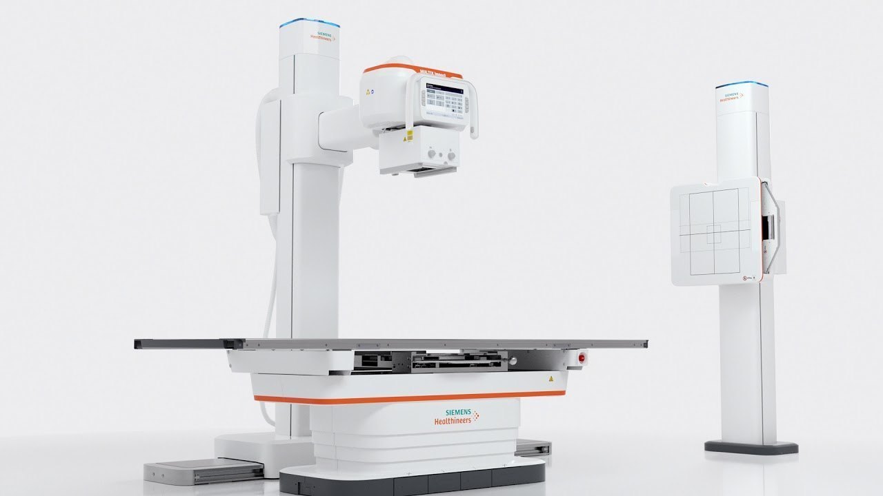
Overview of X-ray - Other Specialized Scans
Beyond the commonly discussed X-ray examinations of the chest, spine, and major joints, X-ray imaging remains a foundational diagnostic tool for visualizing bones and some soft tissues throughout the rest of the body. These "other" specialized X-ray scans target specific anatomical regions or provide focused views for various clinical indications. Examples include X-rays of the **hand, wrist, ankle, hip, skull, abdomen (plain film), facial bones, or extremities (arm/leg)**. Each of these examinations utilizes a small amount of ionizing radiation to produce static images that highlight bone structures, enabling healthcare providers to assess for fractures, dislocations, infections, tumors, or degenerative changes. They are quick, non-invasive procedures, offering a cost-effective and readily available means to obtain crucial diagnostic information for a wide range of medical conditions affecting different parts of the body.
Why Other Specialized X-ray Scans are Done
Other specialized X-ray scans are performed for a variety of diagnostic purposes tailored to the specific body part being examined. For instance, **hand or wrist X-rays** are common after falls or injuries to check for fractures or dislocations, or to evaluate arthritis. **Ankle or hip X-rays** serve similar purposes for lower limb trauma or chronic pain. A **skull X-ray** might be done after head trauma to look for fractures, though CT scans are often preferred for brain injury. A **plain abdominal X-ray** (KUB - kidney, ureter, bladder) can help detect kidney stones, bowel obstructions, or gas patterns. **Facial bone X-rays** are used to assess for fractures after facial trauma. In general, these X-rays are crucial for quickly identifying bone abnormalities, guiding initial treatment decisions, monitoring healing processes, and sometimes as a preliminary step before more advanced imaging like CT or MRI is considered.
Risks
All X-ray procedures, including these specialized "other" scans, involve exposure to a small amount of ionizing radiation. While this radiation carries a very small theoretical risk of cellular damage that could potentially lead to cancer later in life, the diagnostic benefits derived from obtaining vital clinical information typically far outweigh this minimal risk for most patients. Modern X-ray equipment is meticulously designed to minimize radiation exposure to the patient while ensuring high-quality diagnostic images. The radiation dose from these individual X-rays is generally very low, often comparable to a few days of natural background radiation exposure. It is always critical for **pregnant women** or those who suspect they might be pregnant to inform their doctor and the X-ray technologist before any procedure involving radiation. Although the direct radiation to the fetus may be minimal, protective measures such as lead shielding are always used, or alternative imaging methods may be considered if appropriate, to ensure the safety of the developing fetus. The procedure itself is non-invasive and painless.
How You Prepare
Preparation for "other" specialized X-ray scans is generally straightforward and minimal, but it is tailored to the specific body part being imaged. In most cases, there are **no fasting requirements**, so you can eat and drink normally before your appointment. You typically do not need to discontinue any routine medications. The most important aspect of preparation is to **remove any metal objects** from the area of your body that will be X-rayed, as metal can block the X-rays and create artifacts on the images, obscuring important details. This includes jewelry (rings, watches, bracelets, necklaces, earrings, body piercings), eyeglasses, hairpins, and clothing with metal zippers, buttons, or snaps. You may be asked to change into a hospital gown to ensure unobstructed imaging. It is essential to **inform your doctor and the X-ray technologist if you are pregnant or suspect you might be**, so that appropriate precautions can be taken.
What You Can Expect
Before the Test
Before your specialized X-ray scan, you will typically be asked to remove any clothing or jewelry that contains metal from the specific area of your body that will be examined. This is to prevent interference with the X-ray images. You may be provided with a hospital gown to wear for the duration of the procedure, depending on the body part being imaged. The X-ray technologist will explain the process, answer any questions you may have, and ensure you feel comfortable. It is absolutely crucial to inform the technologist if you are pregnant or think you might be pregnant. In such cases, special precautions, such as the use of lead shielding, will be taken to protect the fetus from radiation exposure, or an alternative imaging study may be considered if appropriate. No other specific preparation, such as fasting, is usually required. The technologist will guide you to the X-ray room and help you position yourself correctly.
During the Test
During a specialized X-ray scan, you will be carefully positioned by a trained X-ray technologist. Depending on the body part being examined (e.g., hand, foot, skull, hip), you may be asked to sit, stand, or lie on an X-ray table. Multiple views are typically taken from different angles to capture comprehensive information about the bones and structures in that region. The technologist will position the body part precisely for each view and may use positioning aids like foam blocks to ensure stability. You will be asked to hold very still for a few seconds during each X-ray exposure to prevent blurring of the image. You will hear a click or buzzing sound as the X-ray machine operates, but you will not feel anything during the X-ray exposure itself. The entire process is generally very quick, usually lasting only a few minutes from entering to leaving the X-ray room.
Results
After your specialized X-ray scan is completed, the images will be sent to a radiologist, a medical doctor specially trained in interpreting X-ray and other imaging studies. The radiologist will carefully examine the bones and visible soft tissues of the scanned area for any fractures, dislocations, signs of infection, tumors, degenerative changes, foreign bodies, or other structural abnormalities. A detailed report of these findings will then be prepared and sent to your referring healthcare provider, usually within a few days. Your doctor will then discuss the results with you, explain what they mean for your symptoms or condition, and recommend any necessary further tests (such as MRI or CT if more detailed soft tissue or complex bone information is needed), treatments, or management plans based on the X-ray findings. In emergency or urgent situations, preliminary results may be communicated to your doctor more quickly.