Contrast Materials and Their Function
In medical imaging, contrast materials, also known as contrast agents or contrast media, play a crucial role in enhancing the diagnostic value of various imaging exams, including x-rays, computed tomography (CT), magnetic resonance imaging (MRI), and fluoroscopy. These materials temporarily alter how imaging tools interact with the body, thereby improving the visibility and differentiation of specific structures within the body.
Contrast materials are not permanent dyes; rather, they are substances that modify the appearance of internal organs and tissues on imaging scans. They work by temporarily changing the way x-rays or other imaging modalities interact with different body structures, which helps to highlight and distinguish areas of interest from surrounding tissues. This enhanced contrast allows physicians to better diagnose and evaluate medical conditions.
Contrast materials can be introduced into the body through several methods, including oral ingestion, rectal administration, intravenous injection, or injection into body cavities. For instance, iodine-based and barium-sulfate compounds are commonly used in x-ray and CT imaging. Iodine-based contrasts are injected into veins or arteries, or administered into body cavities, and they work by blocking x-rays, thereby making certain structures appear more prominent on the images. Barium-sulfate is typically used for oral or rectal imaging and is available in various forms, such as powders mixed with water, liquids, pastes, or tablets.
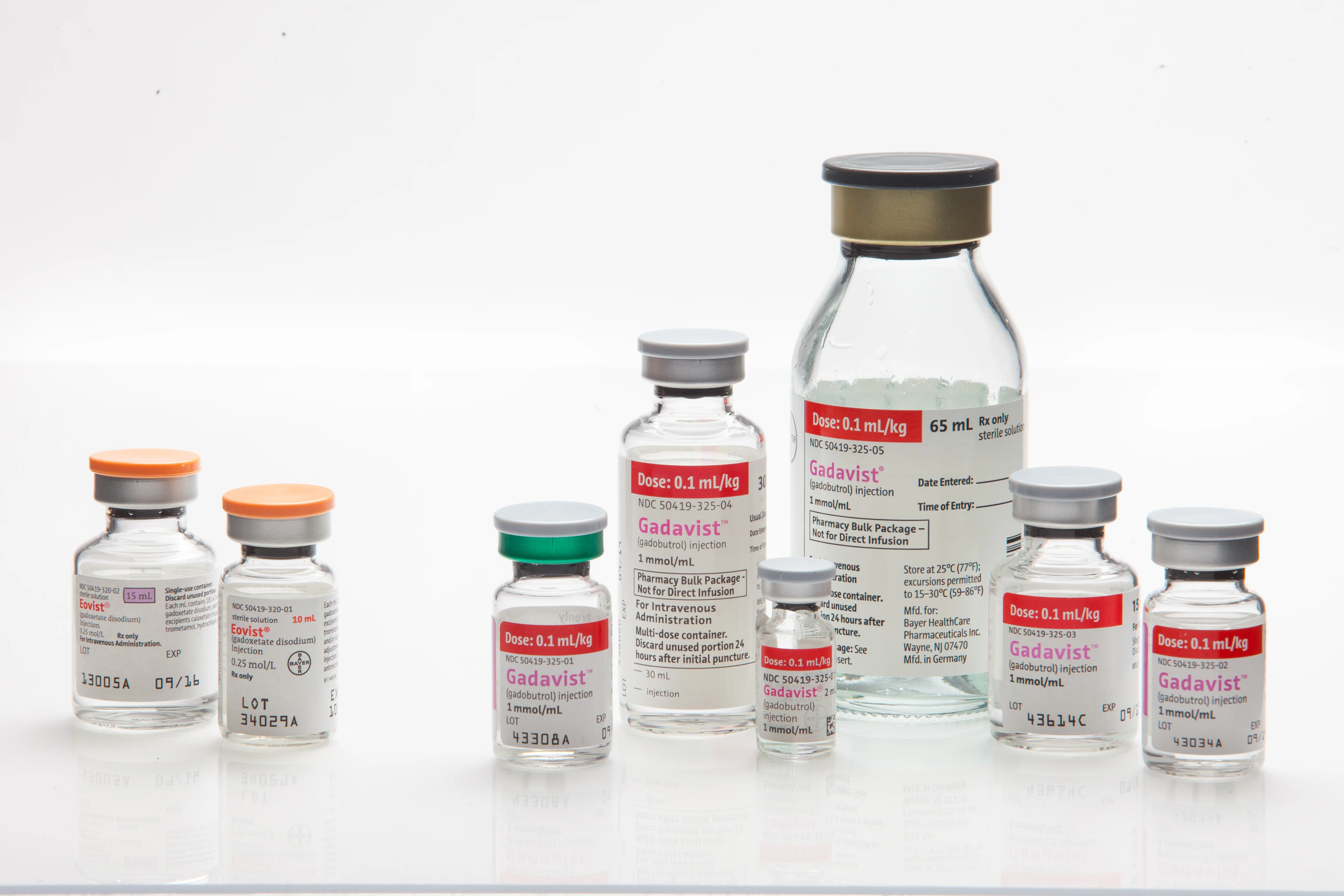
For magnetic resonance imaging (MRI), gadolinium is the primary contrast material. It alters the magnetic properties of nearby water molecules, which enhances the visibility of organs and blood vessels on MRI scans. In addition, saline (salt water) and gases like air are used in certain imaging techniques, while microbubbles and microspheres are utilized in ultrasound imaging, particularly for cardiac evaluations.
After an imaging exam, the contrast materials are either absorbed by the body or expelled through urine or bowel movements. By enhancing the contrast in images, these materials help medical professionals to accurately diagnose and assess various conditions, making them an indispensable tool in modern diagnostic imaging.
Which imaging exams use contrast materials?
Contrast materials are integral to various imaging exams, enhancing the clarity and diagnostic value of the results. Different types of contrast materials are used based on the imaging technique and the area being examined.
Oral contrast materials, such as barium-sulfate, are ingested to improve the visibility of the gastrointestinal (GI) tract during x-rays, fluoroscopy, and CT scans. This method helps to visualize structures including the pharynx, esophagus, stomach, small intestine, and large intestine. In some cases, iodine-based contrast materials may be used instead of barium-sulfate for oral administration.
Rectal contrast materials involve the use of barium-sulfate administered by enema to enhance imaging of the lower GI tract, including the colon and rectum. Iodine-based contrast materials may also be used rectally in certain situations.
Intravenous contrast materials include iodine-based and gadolinium-based agents. Iodine-based contrast is commonly injected into veins to enhance x-ray and CT images, and it is also used during angiograms to visualize arteries. Gadolinium-based contrast is used for enhancing MRI images. These agents improve the visibility of internal organs, such as the brain, heart, lungs, liver, kidneys, and other structures, including the gastrointestinal tract and blood vessels.
Microbubble contrast materials are utilized in ultrasound imaging to enhance the visibility of blood flow and tissue abnormalities. These tiny bubbles, which are smaller than red blood cells, are filled with a gas and reflect ultrasound waves more effectively than surrounding tissues. Microbubbles dissolve within minutes, and the gas is expelled from the body through exhalation. This method is particularly useful for patients with kidney issues or allergies to other contrast agents. Microbubble contrast can be targeted or untargeted; untargeted contrast enhances general blood flow imaging, while targeted contrast binds to specific molecules to highlight particular tissues or abnormalities.
Microbubble contrast-enhanced ultrasound is employed to assess various conditions, including blood perfusion, thrombosis, heart abnormalities, liver and kidney masses, inflammatory activity in bowel diseases, and responses to chemotherapy. This technique offers a radiation-free alternative to other imaging modalities and provides detailed information on the targeted areas of interest.
How safe are contrast materials?
Contrast materials are generally considered safe and are used extensively to enhance the diagnostic quality of imaging exams. They are classified as drugs and, while adverse reactions can occur, most are mild and temporary. Severe reactions are rare but can happen. Radiology departments are well-prepared to handle any serious allergic or other adverse reactions effectively. The safety protocols in place ensure that any potential risks are managed promptly, minimizing the likelihood of serious complications. Overall, the benefits of using contrast materials to obtain clearer and more accurate diagnostic information usually outweigh the risks.
How should I prepare for my imaging procedure with contrast material?
Before your imaging procedure involving contrast material, you will receive detailed instructions tailored to your specific exam. It's important to follow these instructions closely to ensure your safety and the effectiveness of the procedure. Since contrast materials carry a small risk of allergic or adverse reactions, informing your doctor about any of the following conditions is crucial: previous allergic reactions to contrast materials, allergies to foods, drugs, dyes, preservatives, or animals, and a history of heart disease, diabetes, kidney disease, or thyroid problems. These factors can influence the preparation and management strategies recommended for your procedure, helping to mitigate potential risks and optimize the outcomes of your imaging exam.
Side effects and adverse and allergic reactions
When using barium sulfate contrast materials, patients might experience mild side effects such as stomach cramps, diarrhea, nausea, vomiting, or constipation. If these symptoms persist or worsen, it is important to inform your doctor. Immediate medical attention is needed if you experience severe reactions such as hives, itching, red skin, swelling of the throat, difficulty breathing or swallowing, hoarseness, rapid heartbeat, or a bluish skin color. Certain conditions, like cystic fibrosis, severe dehydration, or existing intestinal blockages, can increase the risk of adverse reactions to barium sulfate.
For iodine-based contrast materials, delayed reactions, including rashes that can appear hours to days after the exam, are possible but generally mild. Severe reactions are rare but can include symptoms such as difficulty breathing, swelling of the throat or other body parts, and profound low blood pressure. Common mild to moderate side effects include nausea, vomiting, headache, itching, flushing, and mild skin rash or hives. More severe reactions could involve wheezing, severe skin rash, abnormal heart rhythms, and shortness of breath. Patients with impaired kidney function must be carefully evaluated before receiving iodine-based contrast materials, as they are at higher risk of contrast-induced acute kidney injury.
In MRI scans, gadolinium-based contrast materials are used and are less likely to cause allergic reactions compared to iodine-based agents. However, rare allergic reactions may occur, presenting as hives or itchy eyes, typically manageable with medication. A rare but serious condition, nephrogenic systemic fibrosis (NSF), can affect patients with severe kidney disease, characterized by thickening of the skin and other tissues. Additionally, while gadolinium is generally considered safe, there is evidence of trace retention in various organs, including the brain. This has no known negative effects but might influence the choice of contrast agent, especially in patients undergoing multiple MRI scans.
What will I experience before and after receiving contrast material?
Before undergoing an imaging exam with contrast material, there are specific preparatory steps you might need to follow based on the type of contrast used. If barium sulfate contrast is administered orally or rectally, you may be instructed to fast for a few hours before the exam. For rectal administration, a cleansing regimen involving a special diet and possibly an enema may be required. When swallowing the barium sulfate, the taste might be mildly unpleasant, but it is generally tolerable. If administered rectally, you might experience a sense of abdominal fullness and an urgent need to expel the liquid, though this discomfort is temporary.
Post-exam, it is advisable to drink plenty of fluids to help flush the barium sulfate from your system. Expect bowel movements to appear white for a few days, and some changes in bowel habits may occur within the first 24 hours.
For iodine-based contrast material, which is injected into the bloodstream, you might feel a warm sensation and a metallic taste in your mouth briefly. There may be some discomfort from the needle insertion and potential bruising afterward. Increasing fluid intake post-exam can aid in eliminating the contrast material from your body.
When gadolinium-based contrast material is used, you will typically feel a cool sensation at the injection site, often in your arm, for a minute or two. Discomfort from the needle and possible bruising at the injection site are common. As with other contrast materials, drinking plenty of fluids after the exam helps in the removal of the contrast from your body.
In all cases, if you have not been sedated, there is no need for a recovery period. You can resume your normal activities and diet immediately after the imaging exam.
Pregnancy and contrast materials
Before undergoing any imaging exam with contrast materials, it is crucial for women to inform their physician or radiologic technologist if there is any possibility of pregnancy. This precaution helps to minimize potential risks to the developing fetus. For CT imaging that uses iodinated contrast agents, there is no known significant risk to either the mother or the baby. Nevertheless, discussing any concerns with the radiologist can provide a clearer understanding of the risks and benefits associated with the contrast-enhanced scan.
In the case of MRI, gadolinium-based contrast materials are typically avoided during pregnancy due to unknown risks to the fetus. However, gadolinium may be used if critical information necessary for the mother's health can only be obtained through its use.
For breastfeeding mothers who receive intravenous contrast materials, such as iodine-based or gadolinium-based agents, manufacturers generally recommend not breastfeeding for 24 to 48 hours following the administration of the contrast. This recommendation is based on concerns about the potential effects of contrast agents in breast milk. However, both the American College of Radiology (ACR) and the European Society of Urogenital Radiology have reviewed the data and find no evidence suggesting that the small amounts of contrast agents present in breast milk pose a significant risk. The ACR's Manual on Contrast Media indicates that it is generally safe to continue breastfeeding after receiving gadolinium contrast, but mothers who are concerned may choose to temporarily abstain from breastfeeding for 24 hours and use previously expressed milk during this period.
For detailed information, it is advisable to consult the ACR Manual on Contrast Media and its references available on the ACR website.